Categoria: congresso-2025

  • Effetti di una mobilizzazione molto precoce rispetto che precoce sugli outcome funzionali e psicosociali in fase acuta dopo protesi d’anca

    Effects of very early versus early mobilization and walking on functional and psychosocial outcomes in acute phase after hip arthroplasty

    Autori

    De Leo Davide [Physiotherapy Unit, IRCCS Humanitas Research Hospital, Rozzano, Milan, Italy]

    Temporiti Federico [Department of Biomedical Sciences, Humanitas University, Pieve Emanuele, Milan, Italy] [Physiotherapy Unit, IRCCS Humanitas Research Hospital, Rozzano, Milan, Italy]

    Della Gatta Sofia [Physiotherapy Unit, IRCCS Humanitas Research Hospital, Rozzano, Milan, Italy]

    Conti Davide [Physiotherapy Unit, IRCCS Humanitas Research Hospital, Rozzano, Milan, Italy]

    Adamo Paola [Department of Biomedical Sciences, Humanitas University, Pieve Emanuele, Milan, Italy] [Physiotherapy Unit, IRCCS Humanitas Research Hospital, Rozzano, Milan, Italy]

    Gatti Roberto [Department of Biomedical Sciences, Humanitas University, Pieve Emanuele, Milan, Italy] [Physiotherapy Unit, IRCCS Humanitas Research Hospital, Rozzano, Milan, Italy]

    Background and aims

    Early mobilization and walking after total hip arthroplasty (THA) facilitate an early achievement of discharge criteria1, but heterogeneity in terms of administration timing has been reported2. In fact, mobilization and walking may be delivered in the first postoperative hours (very early) or between 12 and 24 hours (early) after surgery. The study aimed to investigate the effects of very early versus early mobilization and walking on functional and psychosocial outcomes in the acute phase after THA.

    Methods

    In this observational study, 237 patients who underwent THA performed either very early (n=174) or early (n=63) mobilization and walking. Functional mobility (Timed Up and Go – TUG) and pain (Numeric Pain Rating Scale – NPRS) were assessed preoperatively (T0), on the first (T1), and third (T2) postoperative days. Fear of movement (Tampa Scale of Kinesiophobia – TSK) and anxiety (Visual Analogue Scale for anxiety – VAS-A) were also investigated at T0 and T2. Finally, percentage of participants who achieved independent walking with crutches at T1, length of stay (LOS), rate of adverse events, and satisfaction (Visual Analogue Patient Satisfaction Scale – VAPSS) were collected.

    Results

    TUG worsened after surgery and improved from T1 to T2. Pain levels decreased from T1 to T2, while TSK and VAS-A improved from baseline to T2 without significant differences between groups. Similarly, no between-group differences were found for the percentage of patients achieving independent walking at T1, LOS, adverse events rate, and VAPSS.

    Conclusion

    Very early mobilization and walking were not superior to early mobilization and walking on functional and psychosocial outcomes after THA. These results suggest that mobilization and walking may be administered either within the first postoperative hours or on the first postoperative day, increasing its applicability and facilitating the distribution of rehabilitative activities in acute phase after THA.

    REFERENCES

    1. Wainwright TW, Gill M, McDonald DA, et al. Consensus statement for perioperative care in total hip replacement and total knee replacement surgery: Enhanced Recovery After Surgery (ERAS®) Society recommendations. Acta Orthop. 2020;91(1):3-19. doi:10.1080/17453674.2019.1683790
    2. Zhang Q, Chen Y, Li Y, et al. Enhanced recovery after surgery in patients after hip and knee arthroplasty: a systematic review and meta-analysis. Postgrad Med J. 2024;100(1181):159-173. doi:10.1093/postmj/qgad125
  • Avanzamento delle terapie riabilitative attraverso la terapia integrata per l’ictus nella realtà virtuale (ARTIST): training cognitivo e motorio a doppio compito per il recupero post-ictus

    Advancing Rehabilitation Therapies through Integrated Stroke Treatment in Virtual Reality (ARTIST): Cognitive and Motor Dual-Task Training for Post-Stroke Recovery

    Autori

    1Martina Regazzetti, 1Marta Albani Rocchetti, 1Sara Federico, 1Pawel Kiper 1 Healthcare Innovation Technologies Laboratory, IRCCS San Camillo, Venice, Italy

    Background and aims

    • Upper limb deficits are common and disabling in post-stroke patients, with around 80% experiencing motor impairments during the acute phase. Six months post-stroke, up to 60% still face functional difficulties, negatively impacting autonomy and quality of life. As the population ages and the number of chronic patients rises, the demand for rehabilitation services grows. In this context, innovative organizational models and the integration of advanced technologies such as virtual reality (VR) are emerging to enhance rehabilitation effectiveness and efficiency. Motor-cognitive dual task exercises show promise in promoting neuroplasticity and transferring learned skills to daily activities. The primary objective is to evaluate the clinical effectiveness of a motor-cognitive dual task rehabilitation intervention delivered in VR to post-stroke patients with upper limb deficits, comparing two organizational models (1:1 and 2:1) to conventional therapy.
      Secondary objectives include analyzing changes in brain activation and muscle synergies in the experimental groups versus the control group,

    Methods

    This study is a randomized controlled clinical trial to be conducted at IRCCS San Camillo Hospital in Venice, Italy, enrolling 54 post-stroke patients. Participants will be randomly assigned to three groups: a 1:1 experimental group receiving motor-cognitive VR therapy with one dedicated physiotherapist; a 2:1 experimental group with one therapist supervising two patients simultaneously; and a control group receiving conventional upper limb therapy, matched for intensity and duration. Experimental sessions will occur in the SMART room, a sensor-equipped environment developed in the European HosmartAI project, using VRRS Evo systems (Khymeia S.r.l., Padua, Italy). Each session will last one hour, five days a week for three weeks, including progressively tailored motor and cognitive exercises. Visual and auditory feedback will support learning and motivation. Evaluations will occur at baseline and after the intervention, using the following outcome measures: motor function (Fugl-Meyer Assessment for Upper Extremity, Box and Block Test), cognitive function (Mini-Mental State Examination, Oxford Cognitive Screen), neurophysiology (surface electromyography, electroencephalogram), and user experience (System Usability Scale).

    Results

    It is expected that patients in the experimental groups will show clinically and statistically significant improvements compared to the control group in both motor function and neurophysiological activation.

    Conclusion

    The 2:1 organizational model is anticipated to be non-inferior to the 1:1 model, offering a It sustainable and effective approach for post-stroke rehabilitation, particularly in settings with limited professional resources. The analysis of muscle synergies and brain activity will enhance understanding of neuroplastic mechanisms underlying functional recovery and support the development of personalized, technology-enhanced rehabilitation strategies.


    REFERENCES

    Kwakkel, G.; Kollen, B.J.; van der Grond, J.; Prevo, A.J.H. Probability of regaining dexterity in the flaccid upper limb – Impact of severity of paresis and time since onset in acute stroke. Stroke 2003, 34, 2181-2186, doi:10.1161/01.Str.0000087172.16305.Cd.

    1. Hayward, K.S.; Kramer, S.F.; Dalton, E.J.; Hughes, G.R.; Brodtmann, A.; Churilov, L.; Cloud, G.; Corbett, D.; Jolliffe, L.; Kaffenberger, T.; et al. Timing and Dose of Upper Limb Motor Intervention After Stroke: A Systematic Review. Stroke 2021, 52, 3706-3717, doi:10.1161/STROKEAHA.121.034348.
    2. Sathian, K.; Buxbaum, L.J.; Cohen, L.G.; Krakauer, J.W.; Lang, C.E.; Corbetta, M.; Fitzpatrick, S.M. Neurological principles and rehabilitation of action disorders: common clinical deficits. Neurorehabil Neural Repair 2011, 25, 21S-32S, doi:10.1177/1545968311410941.
  • Exoskeleton-Assisted Gait: A Case Series Exploring New Rehabilitation Perspectives in Degenerative Spinal Cord Injury

    Deambulazione Assistita da esoscheletro: una serie di casi che esplora nuove prospettive riabilitative nelle lesioni midollari degenerative

    Autori

    Martina Regazzetti [Healthcare Innovation Technology Lab, IRCCS San Camillo Hospital, Venice, Italy]

    Marta Albani Rocchetti [Healthcare Innovation Technology Lab, IRCCS San Camillo Hospital, Venice, Italy]

    Lazzaro Giovanni [IRCCS San Camillo Hospital, Venice, Italy]

    Federico Sara [Healthcare Innovation Technology Lab, IRCCS San Camillo Hospital, Venice, Italy]

    Cieslik Blazej [Healthcare Innovation Technology Lab, IRCCS San Camillo Hospital, Venice, Italy]

    Kiper Pawel [Healthcare Innovation Technology Lab, IRCCS San Camillo Hospital, Venice, Italy]

    Introduction to clinical case

    Recovery following incomplete spinal cord injury (iSCI) remains challenging, with conventional rehabilitation often emphasizing compensation over functional restoration. As most new SCI cases preserve some motor or sensory pathways, there is increasing interest in therapies that harness neuroplasticity. Robotic exoskeletons provide a promising means to deliver task-specific, repetitive gait training that may promote adaptive neural reorganization. This case series investigates the effects of exoskeleton-assisted walking on motor performance and neuromuscular coordination in individuals with degenerative iSCI.

    Methods

    Two cooperative male patients (aged 77 and 74) with incomplete, non-traumatic SCI participated in a four-week intervention involving one hour of neuromotor physiotherapy followed by one hour of exoskeleton-assisted gait training, three times per week. Functional performance was assessed using the 10-Meter Walk Test. Gait quality and neuromuscular control were further evaluated via spatiotemporal gait analysis and dynamic surface electromyography (sEMG) during overground walking

    Results

    Subject A demonstrated a clinically meaningful increase in walking speed (+1.5 m/s), improved gait symmetry, and more phase-specific activation of key lower limb muscles, including the tibialis anterior, gastrocnemius, and quadriceps. Subject B showed stable walking speed with persistent asymmetries in sEMG patterns, marked by prolonged and compensatory muscle activity, indicating limited neuromuscular adaptation.

    Discussion and clinical relevance

    Given the small number of participants, the current data do not allow us to determine if training with the exoskeleton can improve motor performance during walking in terms of endurance and speed. More data are needed to define in greater detail the impact that using this device can have on walking. Additionally, it is essential to verify on a larger number of patients whether the observed trends are consistent and significant. Examining the specific mechanisms by which the exoskeleton influences muscle activity could provide valuable insights. Comprehensive research with larger cohorts and extended follow-up periods is crucial to fully understand and optimize the potential of exoskeleton-assisted rehabilitation for improving patient outcomes. These findings suggest that robotic exoskeleton training may serve as an effective rehabilitation tool for selected patients with degenerative iSCI. The observed differences in outcomes underscore the importance of individualized approaches and highlight the need for further research into factors influencing responsiveness to robotic interventions.

    REFERENCES

    • Devivo MJ. Epidemiology of traumatic spinal cord injury: trends and future implications. Spinal Cord. 2012 May;50(5):365-72. Epub 2012 Jan 24. PMID: 22270188.
    • Kirshblum SC, Burns SP, Biering-Sorensen F, Donovan W, Graves DE, Jha A, Johansen M, Jones L, Krassioukov A, Mulcahey MJ, Schmidt-Read M, Waring W. International standards for neurological classification of spinal cord injury. J Spinal Cord Med. 2011 Nov;34(6):535-46. PMID: 22330108
    • Gizzi, L., Nielsen, J. F., Felici, F., Ivanenko, Y. P., and Farina, D. (2011). Impulses of activation but not motor modules are preserved in the locomotion of subacute stroke patients. Neurophysiol. 106, 202–210.
    • Sczesny-Kaiser M, Trost R, Aach M, Schildhauer TA, Schwenkreis P, Tegenthoff M. A Randomized and Controlled Crossover Study Investigating the Improvement of Walking and Posture Functions in Chronic Stroke Patients Using HAL Exoskeleton – The HALESTRO Study. Front Neurosci. 2019 Mar 29;13:259. PMID: 30983953;


  • VALUTAZIONE DELL’IMPATTO DELLA STIMOLAZIONE CEREBRALE NON INVASIVA NEI DISTURBI POSTURALI IN PAZIENTI CON MALATTIA DI PARKINSON: UNA REVISIONE SISTEMATICA DELLA LETTERATURA

    VALUTAZIONE DELL’IMPATTO DELLA STIMOLAZIONE CEREBRALE NON INVASIVA NEI DISTURBI POSTURALI IN PAZIENTI CON MALATTIA DI PARKINSON: UNA REVISIONE SISTEMATICA DELLA LETTERATURA

    THE IMPACT OF NON-INVASIVE BRAIN STIMULATION ON POSTURAL DISORDERS IN PATIENTS WITH PARKINSON’S DISEASE: A SYSTEMATIC REVIEW

    Autori

    Martina Ceriani (1Bachelor degree Physiotherapy, School of Medicine, University of Insubria, Varese, Italy 2 Division of Physiotherapy, IRCCS Istituto Auxologico Italiano, Piancavallo (Verbania), Italy.)

    Elisa Ravizzotti (Department of Neurology and Laboratory of Neuroscience, IRCCS Istituto Auxologico Italiano, Milan, Italy. 4 Department of Neuroscience, Rehabilitation, Ophthalmology, Genetics, Maternal and Child Health (DINOGMI), University of Genoa, Genoa, Italy.)

    Background and aims

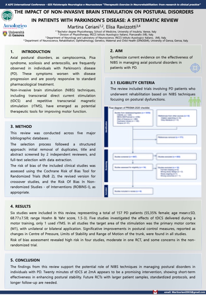
    Axial postural disorders, as camptocormia, Pisa syndrome, scoliosis and anterocollis, are frequently observed in individuals with Parkinson’s disease. These symptoms worsen with disease progression and are poorly responsive to standard pharmacological treatments, leading to a greater functional impairment and a significant decline in quality of life. NIBS techniques, including tDCS and  rTMS, have emerged as potential therapeutic tools for improving motor function.The main purpose of this systematic review was to synthesize current evidence on the effectiveness of NIBS in managing axial postural disorders in patients with PD.

    Methods

    The literature search for this review was conducted across five major bibliographic databases: PubMed, the Cochrane Library, Scopus, Web of Science, and CINAHL. The search strategy, from inception to April 2024, was structured using the Problem-Intervention-Outcome. The selection process followed a structured approach: initial removal of duplicates, title and abstract screened by 2 independent reviewers, and full-text selection with data extraction. The review included trials involving adult patients with PD who underwent rehabilitation interventions based on NIBS techniques. To be eligible, studies had to assess at least one outcome related to postural alignment or postural control, in static or dynamic condition. Studies were excluded if they involved patients with a DBS implant.The risk of bias of the included clinical trial studies was assessed using the Cochrane RoB 2, the  version for crossover studies, and ROBINS-I.

    Results

    From 1.018 records, a total of four RCTs, one cross-over study and one non-randomized clinical trial study, met the inclusion criteria, involving 157 participants with PD at a mild to moderate stage. Five studies investigated the effects of tDCS, only one utilized rTMS. In all studies the target of the intervention was M1, with unilateral or bilateral application and administration in an online setting. The control interventions showed a sham stimulation. Improvements in postural control measures reported as changes in Centre of Pressure, Limits of Stability and Range of Motion of the trunk were found in all studies. Risk of bias assessment revealed high risk in four studies, moderate in one, and some concerns in another one.

    Conclusion

    The findings from this review support the potential role of NIBS techniques in managing postural disorders in individuals with PD. In particular, tDCS appears to be a promising intervention, showing short-term effectiveness in enhancing postural stability. There were limitations such as small sample sizes and short follow-up periods. Future RCTs with larger patient samples, standardized protocols, and longer follow-up are needed.

    REFERENCES

    World Health Organization. who.int. 2023. Parkinson disease.

    Tolosa E, Garrido A, Scholz SW, Poewe W. Challenges in the diagnosis of

    Parkinson’s disease. Vol. 20, The Lancet Neurology. Lancet Publishing Group;

    2021. pag. 385–97.

    Feigin VL, Nichols E, Alam T, Bannick MS, Beghi E, Blake N, et al. Global,

    regional, and national burden of neurological disorders, 1990–2016: a systematic

    analysis for the Global Burden of Disease Study 2016. Lancet Neurol. 1 maggio

    2019;18(5):459–80.

    Vos T, Lim SS, Abbafati C, Abbas KM, Abbasi M, Abbasifard M, et al. Global

    burden of 369 diseases and injuries in 204 countries and territories, 1990–2019: a

    systematic analysis for the Global Burden of Disease Study 2019. The Lancet

    [Internet]. ottobre 2020;396(10258):1204–22. Disponibile su:

    https://linkinghub.elsevier.com/retrieve/pii/S0140673620309259

    Xu T, Dong W, Liu J, Yin P, Wang Z, Zhang L, et al. Disease burden of

    Parkinson’s disease in China and its provinces from 1990 to 2021: findings from

    the global burden of disease study 2021. Lancet Reg Health West Pac. 1 maggio

    2024;46.

    Ben-Shlomo Y, Darweesh S, Llibre-Guerra J, Marras C, San Luciano M, Tanner

    C. The epidemiology of Parkinson’s disease. Vol. 403, The Lancet. Elsevier B.V.;

    2024. pag. 283–92.

    Jankovic J, Tan EK. Parkinson’s disease: Etiopathogenesis and treatment. J

    Neurol Neurosurg Psychiatry. 1 agosto 2020;91(8):795–808.

    Gayed I, Joseph U, Fanous M, Wan D, Schiess M, Ondo W, et al. The Impact of

    DaTscan in the Diagnosis of Parkinson Disease. 2015.

    Marsili L, Rizzo G, Colosimo C. Diagnostic criteria for Parkinson’s disease:

    From James Parkinson to the concept of prodromal disease. Vol. 9, Frontiers in

    Neurology. Frontiers Media S.A.; 2018.

    Margaret M. Hoehn M and MDYM. Parkinsonism Onset, progression, and

    mortality. Neurology Journals. 1998;427–42.11. 12. 13. 14. 15. 16. 17. 18. 19. 20. Movement Disorder Society Task Force report on the Hoehn and Yahr staging

    scale: Status and recommendations The Movement Disorder Society Task Force

    on rating scales for Parkinson’s disease | Enhanced Reader. 2004;

    Goetz CG, Tilley BC, Shaftman SR, Stebbins GT, Fahn S, Martinez-Martin P, et

    al. Movement Disorder Society-Sponsored Revision of the Unified Parkinson’s

    Disease Rating Scale (MDS-UPDRS): Scale presentation and clinimetric testing

    results. Movement Disorders. 15 novembre 2008;23(15):2129–70.

    Goetz CG, Poewe W, Rascol O, Sampaio C, Stebbins GT, Counsell C, et al.

    Movement Disorder Society Task Force report on the Hoehn and Yahr staging

    scale: Status and recommendations. Movement Disorders. settembre

    2004;19(9):1020–8.

    Skorvanek M, Martinez-Martin P, Kovacs N, Rodriguez-Violante M et al.

    Differences in MDS-UPDRS Scores Based on Hoehn and Yahr Stage and

    Disease Duration. Mov Disord Clin Pract. 2017 Mar 11;4(4):536-544..

    Chakra G. A Review of Parkinson’s Disease: Diagnosis and Treatment. Future J

    Pharm Health Sci. 2023;3(4):416–25.

    Leite Silva ABR, Gonçalves de Oliveira RW, Diógenes GP, de Castro Aguiar

    MF, Sallem CC, Lima MPP, et al. Premotor, nonmotor and motor symptoms of

    Parkinson’s Disease: A new clinical state of the art. Vol. 84, Ageing Research

    Reviews. Elsevier Ireland Ltd; 2023.

    Cheung TCK, Schmuckler MA. Multisensory postural control in adults:

    Variation in visual, haptic, and proprioceptive inputs. Hum Mov Sci. 1 ottobre

    2021;79.

    Ivanenko Y, Gurfinkel VS. Human postural control. Vol. 12, Frontiers in

    Neuroscience. Frontiers Media S.A.; 2018.

    Carini F, Mazzola M, Fici C, Palmeri S, Messina M, Damiani P, et al. Posture and

    posturology, anatomical and physiological profiles: Overview and current state

    of art. Vol. 88, Acta Biomedica. Mattioli 1885 S.p.A.; 2017. pag. 11–6.

    Fukunaga JY, Quitschal RM, Doná F, Ferraz HB, Ganança MM, Caovilla HH.

    Postural control in Parkinson’s disease. Braz J Otorhinolaryngol.

    2014;80(6):508–14.21. 22. 23. 24. 25. 26. 27. 28. 29. 30. Robinson K, Roalf DR, Cianci H, Bunting-Perry L, Dennison A, Roalf D, et al.

    Falling risk factors in Parkinson’s disease. Vol. 20, NeuroRehabilitation. IOS

    Press; 2005.

    Allcock LM, Rowan EN, Steen IN, Wesnes K, Kenny RA, Burn DJ. Impaired

    attention predicts falling in Parkinson’s disease. Parkinsonism Relat Disord.

    febbraio 2009;15(2):110–5.

    Sarter M, Albin RL, Kucinski A, Lustig C. Where attention falls: Increased risk

    of falls from the converging impact of cortical cholinergic and midbrain

    dopamine loss on striatal function. Vol. 257, Experimental Neurology. Academic

    Press Inc.; 2014. pag. 120–9.

    Crouse JJ, Phillips JR, Jahanshahi M, Moustafa AA. Postural instability and falls

    in Parkinson’s disease. Vol. 27, Reviews in the Neurosciences. Walter de Gruyter

    GmbH; 2016. pag. 549–55.

    Paul SS, Sherrington C, Canning CG, Fung VSC, Close JCT, Lord SR. The

    relative contribution of physical and cognitive fall risk factors in people with

    Parkinson’s disease: A large prospective cohort study. Neurorehabil Neural

    Repair. marzo 2014;28(3):282–90.

    Borel L, Alescio-Lautier B. Posture and cognition in the elderly: Interaction and

    contribution to the rehabilitation strategies. Vol. 44, Neurophysiologie Clinique.

    2014. pag. 95–107.

    Phenotype of postural instability:gait difficulty in Parkinson disease: relevance to

    cognitive impairment and mechanism relating pathological proteins and

    neurotransmitters | Enhanced Reader.

    Visser M, Marinus J, Bloem BR, Kisjes H, van den Berg BM, van Hilten JJ.

    Clinical tests for the evaluation of postural instability in patients with parkinson’s

    disease. Arch Phys Med Rehabil. 2003 Nov;84(11):1669-74.

    Jacobs J V., Horak FB, Tran VK, Nutt JG. Multiple balance tests improve the

    assessment of postural stability in subjects with Parkinson’s disease. J Neurol

    Neurosurg Psychiatry. 2006;77(3):322–6.

    Walter Maetzler 1 JDKSJJFBRB. Quantitative wearable sensors for objective

    assessment of Parkinson’s disease. Mov Disord. 12 settembre 2013;31. 32. 33. 34. 35. 36. 37. 38. 39. 40. Ferreira-Peruzzo Silvia AparecidaORCID Icon CCHFIZMBIRPRMRPI&

    THAGI. Static posturography analysis for postural instability in patients with

    Parkinson’s disease. Int J Neurosci. 2023;

    Ferraris C, Votta V, Nerino R, Chimienti A, Priano L, Mauro A. At-home

    assessment of postural stability in parkinson’s disease: a vision-based approach. J

    Ambient Intell Humaniz Comput. 1 maggio 2024;15(5):2765–78.

    Shirahige L, Leimig B, Baltar A, Bezerra A, de Brito CVF, do Nascimento YSO,

    et al. Classification of Parkinson’s disease motor phenotype: a machine learning

    approach. J Neural Transm. 1 dicembre 2022;129(12):1447–61.

    Abe K, Uchida Y, Notani M. Camptocormia in Parkinson’s disease. Parkinsons

    Dis. 2010 Jun 30;2010:267640.

    Margraf NG, Granert O, Hampel J, Wrede A, Schulz-Schaeffer WJ, Deuschl G.

    Clinical Definition of Camptocormia in Parkinson’s Disease. Mov Disord Clin

    Pract. 2016 Oct 11;4(3):349-357.

    Tinazzi M, Geroin C, Bhidayasiri R, Bloem BR, Capato T, Djaldetti R et al.

    International Parkinson and Movement Disorders Society Task Force on

    Postural Abnormalities. Task Force Consensus on Nosology and Cut-Off Values

    for Axial Postural Abnormalities in Parkinsonism. Mov Disord Clin Pract. 2022

    May 9;9(5):594-603. .

    Furusawa Y, Mukai Y, Kobayashi Y, Sakamoto T, Murata M. Role of the

    external oblique muscle in upper camptocormia for patients with Parkinson’s

    disease. Vol. 27, Movement Disorders. 2012. pag. 802–3.

    Fasano A, Geroin C, Berardelli A, Bloem BR, Espay AJ, Hallett M, Lang AE,

    Tinazzi M. Diagnostic criteria for camptocormia in Parkinson’s disease: A

    consensus-based proposal. Parkinsonism Relat Disord. 2018 Aug;53:53-57.

    Srivanitchapoom P, Hallett M. Camptocormia in Parkinson’s disease: definition,

    epidemiology, pathogenesis and treatment modalities. J Neurol Neurosurg

    Psychiatry. 2016 Jan;87(1):75-85. doi: 10.1136/jnnp-2014-310049. Epub 2015

    Apr 20.

    Tinazzi M, Geroin C, Gandolfi M, Smania N, Tamburin S, Morgante F, Fasano

    A. Pisa syndrome in Parkinson’s disease: An integrated approach from

    pathophysiology to management. Mov Disord. 2016 Dec;31(12).41. 42. 43. 44. 45. 46. 47. 48. 49. 50. 51. Miletic V. Pisa syndrome in Parkinson’s disease: diagnostic and management

    challenges. Research and Reviews in Parkinsonism. 2016;6:29-35.

    Geroin C, Artusi CA, Gandolfi M, Zanolin E, Ceravolo R, Capecci M, et al.

    Does the Degree of Trunk Bending Predict Patient Disability, Motor

    Impairment, Falls, and Back Pain in Parkinson’s Disease? Front Neurol. 31

    marzo 2020;11.

    Tinazzi M, Fasano A, Geroin C, Morgante F, Ceravolo R, Rossi S et al. Italian

    Pisa Syndrome Study Group. Pisa syndrome in Parkinson disease: An

    observational multicenter Italian study. Neurology. 2015 Nov 17;85(20):1769-

    79..

    Vitale C, Falco F, Trojano L, Erro R, Moccia M, Allocca R, et al.

    Neuropsychological correlates of Pisa syndrome in patients with Parkinson’s

    disease. Acta Neurol Scand. 1 agosto 2016;134(2):101–7.

    Cannas A, Solla P, Floris G, Tacconi P, Serra A, Piga M, Marrosu F, Marrosu

    MG. Reversible Pisa syndrome in patients with Parkinson’s disease on

    dopaminergic therapy. J Neurol. 2009 Mar;256(3):390-5.

    Artusi CA, Montanaro E, Tuttobene S, Romagnolo A, Zibetti M, Lopiano L.

    Pisa Syndrome in Parkinson’s Disease Is Associated With Specific Cognitive

    Alterations. Front Neurol. 2019 May 31;10:577.

    Lai Y, Song Y, Su D, Wang L, Zhang C, Sun B, et al. Pallidal stimulation as

    treatment for camptocormia in Parkinson’s disease. NPJ Parkinsons Dis. 1

    dicembre 2021;7(1).

    Chieng LO, Madhavan K, Wang MY. Deep brain stimulation as a treatment for

    Parkinson’s disease related camptocormia. Vol. 22, Journal of Clinical

    Neuroscience. Churchill Livingstone; 2015. pag. 1555–61.

    Ando Y, Fujimoto K ichi, Ikeda K, Utsumi H, Okuma Y, Oka H, et al. Postural

    Abnormality in Parkinson’s Disease: A Large Comparative Study With General

    Population. Mov Disord Clin Pract. 1 marzo 2019;6(3):213–21.

    Balestrino R, Schapira AHV. Parkinson disease. Vol. 27, European Journal of

    Neurology. Blackwell Publishing Ltd; 2020. pag. 27–42.

    Sprenger F, Poewe W. Management of motor and non-motor symptoms in

    parkinson’s disease. Vol. 27, CNS Drugs. 2013. pag. 259–72.52. 53. 54. 55. 56. 57. 58. 59. 60. 61. 62. Hariz M, Blomstedt P. Deep brain stimulation for Parkinson’s disease. Vol. 292,

    Journal of Internal Medicine. John Wiley and Sons Inc; 2022. pag. 764–78.

    N. Bolognini GV. Stimolare il cervello – manuale di stimolazione cerebrale non

    invasiva . Il Mulino, curatore. 2015.

    Kesikburun S. Non-invasive brain stimulation in rehabilitation. Turk J Phys Med

    Rehabil. 2022 Mar 1;68(1):1-8.

    Lefaucheur JP, André-Obadia N, Antal A, Ayache SS, Baeken C, Benninger DH,

    et al. Evidence-based guidelines on the therapeutic use of repetitive transcranial

    magnetic stimulation (rTMS). Vol. 125, Clinical Neurophysiology. Elsevier

    Ireland Ltd; 2014. pag. 2150–206.

    Lefaucheur JP, Aleman A, Baeken C, Benninger DH, Brunelin J, Di Lazzaro V,

    et al. Evidence-based guidelines on the therapeutic use of repetitive transcranial

    magnetic stimulation (rTMS): An update (2014–2018). Vol. 131, Clinical

    Neurophysiology. Elsevier Ireland Ltd; 2020. pag. 474–528.

    Schulz R, Gerloff C, Hummel FC. Non-invasive brain stimulation in

    neurological diseases. Vol. 64, Neuropharmacology. 2013. pag. 579–87.

    Aderinto N, Olatunji G, Muili A, Kokori E, Edun M, Akinmoju O, et al. A

    narrative review of non-invasive brain stimulation techniques in neuropsychiatric

    disorders: current applications and future directions. Vol. 60, Egyptian Journal of

    Neurology, Psychiatry and Neurosurgery. Springer Science and Business Media

    Deutschland GmbH; 2024.

    He W, Fong PY, Leung TWH, Huang YZ. Protocols of non-invasive brain

    stimulation for neuroplasticity induction. Vol. 719, Neuroscience Letters.

    Elsevier Ireland Ltd; 2020.

    Von Bernhardi R, Eugenín-Von Bernhardi L, Eugenín J. What is neural

    plasticity? In: Advances in Experimental Medicine and Biology. Springer New

    York LLC; 2017. pag. 1–15.

    Kania BF, Wrońska D, Zięba D. Introduction to Neural Plasticity Mechanism. J

    Behav Brain Sci. 2017;07(02):41–9.

    Zewdie E, Ciechanski P, Kuo HC, Giuffre A, Kahl C et al. Safety and tolerability

    of transcranial magnetic and direct current stimulation in children: Prospective63. 64. 65. 66. 67. 68. 69. 70. 71. single center evidence from 3.5 million stimulations. Brain Stimul. 2020 May-

    Jun;13(3):565-575..

    Matsumoto H, Ugawa Y. Adverse events of tDCS and tACS: A review. Vol. 2,

    Clinical Neurophysiology Practice. Elsevier B.V.; 2017. pag. 19–25.

    McClintock SM, Reti IM, Carpenter LL, McDonald WM, Dubin M, Taylor SF et

    al. National Network of Depression Centers rTMS Task Group; American

    Psychiatric Association Council on Research Task Force on Novel Biomarkers

    and Treatments. Consensus Recommendations for the Clinical Application of

    Repetitive Transcranial Magnetic Stimulation (rTMS) in the Treatment of

    Depression. J Clin Psychiatry. 2018 Jan/Feb;79(1).

    Page MJ, McKenzie JE, Bossuyt PM, Boutron I, Hoffmann TC, Mulrow CD, et

    al. The PRISMA 2020 statement: An updated guideline for reporting systematic

    reviews. Vol. 372, The BMJ. BMJ Publishing Group; 2021.

    Sterne JAC, Savović J, Page MJ, Elbers RG, Blencowe NS, Boutron I et al. RoB

    2: a revised tool for assessing risk of bias in randomised trials. BMJ. 2019 Aug

    28;

    Revised Cochrane risk-of-bias tool for randomized crossover trials SHORT

    VERSION (CRIBSHEET).

    The Risk Of Bias In Non-randomized Studies-of Interventions (ROBINS-I)

    assessment tool (version for cohort-type studies) ROBINS-I tool (Stage I): At

    protocol stage Specify the review question.

    Yang C, Xu Y, Feng X, Wang B, Du Y, Wang K, Lü J, Huang L, Qian Z, Wang

    Z, Chen N, Zhou J, Zhang C, Liu Y. Transcranial Temporal Interference

    Stimulation of the Right Globus Pallidus in Parkinson’s Disease. Mov Disord.

    2024 Aug 12. .

    Romero JP, Moreno-Verdú M, Arroyo-Ferrer A, Serrano JI, Herreros-Rodríguez

    J, García-Caldentey J, et al. Clinical and neurophysiological effects of bilateral

    repetitive transcranial magnetic stimulation and EEG-guided neurofeedback in

    Parkinson’s disease: a randomized, four-arm controlled trial. J Neuroeng

    Rehabil. 1 dicembre 2024;21(1).

    Beretta VS, Orcioli-Silva D, Zampier VC, Moraca GAG, Pereira MP, Gobbi

    LTB, et al. Eight sessions of transcranial electrical stimulation for postural72. 73. 74. 75. 76. 77. 78. 79. response in people with Parkinson’s disease: A randomized trial. Gait Posture. 1

    ottobre 2024;114:1–7.

    Marcus Grobe-Einsler ALJWOK. RTMS of the Cerebellum Using an

    Accelerated Stimulation Protocol Improved Gait in Parkinson’s Disease.

    Neurorehabil Neural Repair. luglio 2024;539–50.

    TRANSCRANIAL DIRECT CURRENT STIMULATION (t-DCS) AS ADD-

    ON TO NEUROREHABILITATION OF PISA SYNDROME IN

    PARKINSON DISEASE. Clinicaltrials.gov. 1 gennaio 2020;0(0).

    Qi J, KG, SK, SS, MM. The effects of non-invasive transcranial brain current

    stimulation (tDCS) on posture over stable and unstable surfaces in people with

    Parkinson’s: a randomised doubleblind sham-controlled crossover study. J

    Parkinsons Dis. 1 gennaio 2019;9(1):190.

    Kaut O, GEM. Noninvasive brain stimulation improves motor function in

    Parkinson’s disease. Neurologie und rehabilitation. 1 gennaio 2023;23(0).

    Rashid-Lopez R, CGAJ, MGP, SF ez, FL, LSE, SF, CCF, REG, ERR, GRJJ.

    Marked improvement of postural and gait disturbances in Parkinson’s disease

    with bilateral primary motor area intermittent theta-burst stimulation may be

    linked to increased putamen-cortico-cerebellar functional connectivity: a case

    report. Brain stimulation2. 1 gennaio 2023;16(1):271–2.

    Rashid-López R, MGP, CGÁJ, SFFL, CCF, SF et al. Bilateral primary motor

    area intermittent theta-burst stimulation may alleviate gait and postural

    disturbances in Parkinson’s disease patients by astrocytic modulation, caudate

    volume changes, and increased functional neuroplasticity. Parkinsonism Relat

    Disord. 1 gennaio 2024;123(0).

    Radder DLM, Lígia Silva de Lima A, Domingos J, Keus SHJ, van Nimwegen M,

    Bloem BR, de Vries NM. Physiotherapy in Parkinson’s Disease: A Meta-Analysis

    of Present Treatment Modalities. Neurorehabil Neural Repair. 2020

    Oct;34(10):871-880.).

    Legutke BR, Gobbi LTB, Orcioli-Silva D, Santos PCR dos, Moraca GAG,

    Vitório R, et al. Transcranial direct current stimulation suggests not improving

    postural control during adapted tandem position in people with Parkinson’s

    disease: A pilot study. Behavioural Brain Research. 24 agosto 2023;452.80. 81. 82. 83. 84. 85. 86. 87. Beretta VS, Vitório R, Nóbrega-Sousa P, Conceição NR, Orcioli-Silva D, Pereira

    MP, et al. Effect of Different Intensities of Transcranial Direct Current

    Stimulation on Postural Response to External Perturbation in Patients With

    Parkinson’s Disease. Neurorehabil Neural Repair. 1 novembre

    2020;34(11):1009–19.

    Beretta VS, Orcioli-Silva D, Conceição NR, Nóbrega-Sousa P, Pereira MP,

    Gobbi LTB, et al. tDCS application for postural control in Parkinson’s disease:

    Effects are associated with baseline characteristics. Parkinsonism Relat Disord. 1

    dicembre 2021;93:62–5.

    De Icco R, Putortì A, Allena M, Avenali M, Dagna C, Martinelli D, et al. Non-

    Invasive Neuromodulation in the Rehabilitation of Pisa Syndrome in Parkinson’s

    Disease: A Randomized Controlled Trial. Front Neurol. 14 aprile 2022;13.

    Yitayeh A, Teshome A. The effectiveness of physiotherapy treatment on balance

    dysfunction and postural instability in persons with Parkinson’s disease: A

    systematic review and meta-analysis. BMC Sports Sci Med Rehabil. 11 febbraio

    2016;8(1).

    Palheta De Lima K, Nascimento Da Silva C, Ferreira De Seixas N, De M,

    Maneschy S, Lima N, et al. Effect of resistance training on balance and postural

    control in people with Parkinson’s: A systematic review. Vol. 56, Rev Cient Soc

    Esp Enferm Neurol. 2022.

    Coelho DB, de Oliveira CEN, Guimarães MVC, Ribeiro de Souza C, dos Santos

    ML, de Lima-Pardini AC. A systematic review on the effectiveness of

    perturbation-based balance training in postural control and gait in Parkinson’s

    disease. Vol. 116, Physiotherapy (United Kingdom). Elsevier Ltd; 2022. pag. 58–

    71.

    Bath JE, Wang DD. Unraveling the threads of stability: A review of the

    neurophysiology of postural control in Parkinson’s disease. Vol. 21,

    Neurotherapeutics. Elsevier B.V.; 2024.

    Fatemeh Ehsani ASMZRHSJ. The effects of cerebellar transcranial direct current

    stimulation on static and dynamic postural stability in older individuals: a

    randomized double-blind sham-controlled study. European Journal of

    Neuroscience. ottobre 2017;
  • Efficacia della Fisioterapia Psicologicamente Informata in teleriabilitazione per persone con dolore cronico: una revisione sistematica con meta-analisi

    Efficacia della Fisioterapia Psicologicamente Informata in teleriabilitazione per persone con dolore cronico: una revisione sistematica con meta-analisi

    Effectiveness of remotely-delivered Psychologically-Informed Physical Therapy (PIPT) for people with chronic pain: a systematic review with meta-analysis

    Autori

    Matteo Cioeta [Department of Medicine and Health Science “Vincenzo Tiberio”, University of Molise, Campobasso, Italy]

    Luca Giacobbe [Department of Human Neurosciences, Sapienza University of Rome, 00185 Rome, Italy]

    Marco Medugnp [Department of Human Neurosciences, Sapienza University of Rome, 00185 Rome, Italy]

    Davide Lampasi [Department of Human Neurosciences, Sapienza University of Rome, 00185 Rome, Italy]

    Leonardo Pellicciari [IRCCS Istituto delle Scienze Neurologiche di Bologna, Bologna, Italy]

    Giuseppe Giovannico [Department of Medicine and Health Science “Vincenzo Tiberio”, University of Molise, Campobasso, Italy]

    Germano Guerra [Department of Medicine and Health Science “Vincenzo Tiberio”, University of Molise, Campobasso, Italy]

    Giovanni Galeoto [Department of Human Neurosciences, Sapienza University of Rome, 00185 Rome, Italy]

    Background and aims

    To evaluate the effectiveness of remotely-delivered Psychologically-Informed Physical Therapy (PIPT) for individuals with chronic pain (CP) through a systematic review and meta-analysis

    Methods

    Seven databases were searched up to May 2025. Randomized controlled trials (RCTs) assessing the effects of PIPT by physiotherapists for people with CP were included. Primary outcomes were pain and disability; secondary outcomes included quality of life, self-efficacy, kinesiophobia, and costs. The risk of bias was assessed using the Cochrane RoB 2.0 tool. Random effect meta-analyses were performed. GRADE approach was used to rate the certainty of evidence.

    Results

    Four RCTs (n=911 participants) involving patients with knee osteoarthritis were included. The meta-analysis showed low certainty of evidence that remotely-delivered PIPT reduces pain (MD = -0.81 at 6 months; -0.50 at 12 months) and moderate certainty of evidence that it can reduce disability (MD = -3.44 at 6 months; -5.71 at 12 months) compared to other remote interventions. Quality of life improvements were also reported but did not exceed the minimal clinically important difference. One study showed improved self-efficacy but no significant effects on kinesiophobia or healthcare costs. The risk of bias was assessed as ranging from some concerns to high.

    Conclusion

    Remotely delivered PIPT appears effective in improving pain and disability in individuals with osteoarthritis. Although effect sizes were small, these findings support the integration of psychological principles into telerehabilitation. Further research is warranted to explore its generalizability to other chronic pain conditions and to compare its effectiveness against psychologist-led interventions.

    REFERENCES

    1. Treede RD, Rief W, Barke A, Aziz Q, Bennett MI, Benoliel R, Cohen M, Evers S, Finnerup NB, First MB, Giamberardino MA, Kaasa S, Korwisi B, Kosek E, Lavand’homme P, Nicholas M, Perrot S, Scholz J, Schug S, Smith BH, Svensson P, Vlaeyen JWS, Wang SJ. Chronic pain as a symptom or a disease: the IASP Classification of Chronic Pain for the International Classification of Diseases (ICD-11). Pain. 2019 Jan;160(1):19-27. doi: 10.1097/j.pain.0000000000001384. PMID: 30586067.
    2. Main CJ, Steven Z. George, Psychologically Informed Practice for Management of Low Back Pain: Future Directions in Practice and Research, Physical Therapy, Volume 91, Issue 5, 1 May 2011, Pages 820–824, https://doi.org/10.2522/ptj.20110060
    3. Main CJ, Ballengee LA, George SZ, Beneciuk JM, Greco CM, Simon CB Psychologically Informed Practice: The Importance of Communication in Clinical Implementation, Physical Therapy, Volume 103, Issue 7, July 2023, pzad047, https://doi.org/10.1093/ptj/pzad047
    4. Doorley JD, Lentz TA, Yeh GY, Wayne PM, Archer KR, Vranceanu AM. Technology-Enhanced Delivery Models to Facilitate the Implementation of Psychologically Informed Practice for Chronic Musculoskeletal Pain. Phys Ther. 2022 Dec 30;103(1):pzac141. doi: 10.1093/ptj/pzac141. PMID: 36210757; PMCID: PMC10071498.
  • “Ho bisogno di una risonanza?” Esplorazione del perchè i pazienti con dolore cronico lombare non specifico richiedono valutazioni diagnostiche strumentali: uno studio fenomenologico

    “Ho bisogno di una risonanza?” Esplorazione del perchè i pazienti con dolore cronico lombare non specifico richiedono valutazioni diagnostiche strumentali: uno studio fenomenologico

    “Do I need an imaging?” Exploring why patients with non-specific chronic low back pain request diagnostic instrumental evaluation: a phenomenological qualitative study

    Autori

    Graziana Lullo [Department of Medicine and Health Science “Vincenzo Tiberio”, University of Molise, 86100 Campobasso, Italy]

    Gabriele Giannotta [Department of Medicine and Health Science “Vincenzo Tiberio”, University of Molise, 86100 Campobasso, Italy]

    Andrea Tamborrino [Department of Medicine and Health Science “Vincenzo Tiberio”, University of Molise, 86100 Campobasso, Italy]

    Firas Mourad [Department of Physiotherapy, LUNEX International University of Health, Exercise and Sports, Differdange, Luxembourg]

    Massimo Esposto [Department of Medicine and Health Science “Vincenzo Tiberio”, University of Molise, 86100 Campobasso, Italy]

    Giuseppe Giovannico [Department of Medicine and Health Science “Vincenzo Tiberio”, University of Molise, 86100 Campobasso, Italy]

    Morten Hoegh [Department of Health Science and Technology, Faculty of Medicine, Aalborg University, Denmark]

    Matteo Cioeta [Department of Medicine and Health Science “Vincenzo Tiberio”, University of Molise, 86100 Campobasso, Italy]

    Background and aims

    Chronic non-specific low back pain (cLBP) is widely acknowledged to have a multifactorial origin, yet many patients still request advanced imaging—especially magnetic resonance imaging (MRI)—even when clinical guidelines deem it unnecessary. This qualitative, phenomenological study set out to clarify why people with cLBP turn to MRI, exploring the beliefs that shape their expectations and the psychological impact of the results. By illuminating these drivers, the authors aimed to inform patient-centred communication strategies capable of aligning clinical practice with evidence-based recommendations.

    Methods

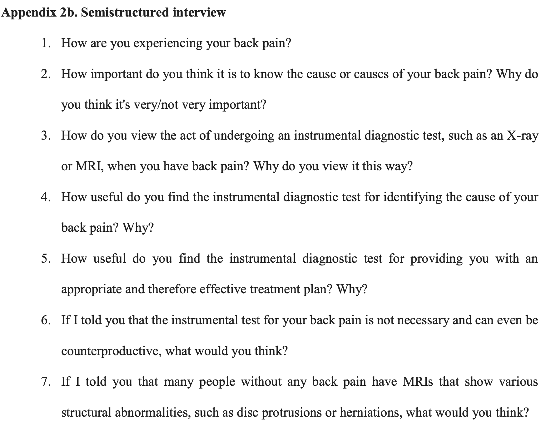
    Eleven adults (six women, five men; mean age = 53 ± 15.7 years) living with cLBP participated in semi-structured, in-depth interviews. Questions probed how each individual interprets pain, perceives the usefulness of imaging, and copes with the findings. Interviews were transcribed verbatim and analysed thematically using an inductive, interpretative approach consistent with phenomenological traditions. Rigour was reinforced through investigator triangulation and member checking, ensuring that emergent themes authentically reflected participants’ lived experiences.

    Results

    Three overarching themes emerged:

    1. Cognitive dissonance: need to ‘know the cause,’ but recognition of multifactorial pain. Participants simultaneously recognised that cLBP rarely stems from a single anatomical lesion and yet remained convinced that a visual confirmation of spinal changes would validate their pain and guide treatment.

    2. Imaging as a treatment guide: seen as useful post-conservative treatment failure; mixed on necessity. Imaging was viewed as an unavoidable milestone once conservative measures (analgesics, physiotherapy, exercise) seemed ineffective. Patients believed that detailed morphology would replace “trial-and-error” management with a more tailored, efficient plan, even though most acknowledged guideline warnings against routine MRI.

    3. Imaging has difference consequences: reassuring for some; anxiety-inducing and misleading for others. Some interviewees felt reassured when MRI identified disc herniations or protrusions, interpreting the images as objective proof of suffering that motivated adherence to rehabilitation. Others reacted with catastrophising thoughts, heightened fear of movement, and a sense of fragility—responses that can undermine functional recovery.


    Conclusion

    Patients’ requests for MRI in cLBP are driven less by ignorance of guidelines and more by a psychological quest for validation, precision, and control. Healthcare professionals should therefore complement evidence-based recommendations with clear, empathetic dialogue about the limited diagnostic and prognostic value of routine imaging. Explaining that structural changes are common in asymptomatic populations, involving patients in shared decision-making, and offering active, lifestyle-oriented alternatives may curb unnecessary imaging, promoting more effective self-management. Enhancing clinicians’ communication skills is thus pivotal to bridge the gap between guidelines and patient expectations in chronic low back pain care.

    REFERENCES

    1. Ferreira, Manuela L et al. Global, regional, and national burden of low back pain, 1990–2020, its attributable risk factors, and projections to 2050: a systematic analysis of the Global Burden of Disease Study 2021 he Lancet Rheumatology, Volume 5, Issue 6, e316 – e329

    2. Oliveira CB, Maher CG, Pinto RZ, Traeger AC, Lin CC, Chenot JF, van Tulder M, Koes BW. Clinical practice guidelines for the management of non-specific low back pain in primary care: an updated overview. Eur Spine J. 2018 Nov;27(11):2791-2803. doi: 10.1007/s00586-018-5673-2. Epub 2018 Jul 3. PMID: 29971708.

    3. Dagenais S, Galloway EK, Roffey DM. A systematic review of diagnostic imaging use for low back pain in the United States. Spine J. 2014 Jun 1;14(6):1036-48. doi:10.1016/j.spinee.2013.10.031.

    4. Pike, A., Patey, A., Lawrence, R., Aubrey-Bassler, K., Grimshaw, J., Mortazhejri, S., Dowling, S., Jasaui, Y., De-implementing Wisely Research Group, & Hall, A. (2022). Barriers to following imaging guidelines for the treatment and management of patients with low-back pain in primary care: a qualitative assessment guided by the Theoretical Domains Framework. BMC primary care, 23(1), 143. https://doi.org/10.1186/s12875-022-01751-6

    5. O’Keeffe M, Ferreira GE, Harris IA, Darlow B, Buchbinder R, Traeger AC, Zadro JR, Herbert RD, Thomas R, Belton J, Maher CG. Effect of diagnostic labelling on management intentions for non-specific low back pain: A randomized scenario-based experiment. Eur J Pain. 2022 Aug;26(7):1532-1545. doi: 10.1002/ejp.1981.

  • Efficacia delle manipolazioni spinali a alta velocità e bassa ampiezza in pazienti con radicolopatia: una revisione sistematica con meta-analisi di studi randomizzati controllati

    Efficacia delle manipolazioni spinali a alta velocità e bassa ampiezza in pazienti con radicolopatia: una revisione sistematica con meta-analisi di studi randomizzati controllati

    Efficacy of spinal high-velocity-low-amplitude thrust manipulations in patients with radiculopathy: a systematic review with meta-analysis of randomized controlled studies

    Autori

    Giuseppe Giovannico [Department of Medicine and Health Science “Vincenzo Tiberio”, University of Molise, Campobasso, Italy]

    Matteo Cioeta [Department of Medicine and Health Science “Vincenzo Tiberio”, University of Molise, Campobasso, Italy]

    Gabriele Giannotta [Associazione “La Nostra Famiglia” – IRCCS "E. Medea" – Unit for Severe disabilities in developmental age and young adults (Developmental Neurology and Neurorehabilitation) – Brindisi, Italy]

    Silvia Bargeri [IRCCS Istituto Ortopedico Galeazzi, Unit of Clinical Epidemiology, Milan, Italy]

    Fabrizio Brindisino [Department of Medicine and Health Science “Vincenzo Tiberio”, University of Molise, Campobasso, Italy]

    Leonardo Pellicciari [IRCCS Istituto delle Scienze Neurologiche di Bologna, Bologna, Italy]

    Background and aims

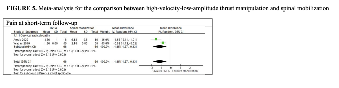
    A multimodal approach, including manual therapy, is the most efficient approach for cervical, thoracic, and lumbar radiculopathy. Among manual therapy techniques spinal high-velocity-low-amplitude thrust (HVLAT) manipulation is widely used. However, the efficacy of HVLAT for cervical, thoracic, and lumbar radiculopathy remains uncertain, and a comprehensive literature review is currently lacking on the HVLAT efficacy on thoracic and lumbar radiculopathy. Therefore, this study aimed to assess the HVLAT efficacy in patients with radiculopathy.

    Methods

    The study protocol was a-priori registered on PROSPERO (CRD42022343394). Medline, CINAHL, EMBASE, CENTRAL, PEDro were searched from their inception until May 2024. Randomized controlled studies assessing the effect of HVLATs in adult patients with cervical, thoracic, or lumbar radiculopathy on pain intensity, disability and other clinical outcomes (range of motion [ROM], quality of life, adverse events) were included. Two independent reviewers performed the study selection, data extrapolation, assessment of risk of bias (Revised Cochrane RoB Tool 2.0), certainty of evidence (CoE) (GRADE approach) and clinical relevance (defined as an additional 20% improvement between groups based on the baseline values of our dataset). If at least two studies assessed the same outcome, meta-analyses using a random-effects model were performed with appropriate effect sizes.

    Results

    Results: Eleven RCTs (N=991), all at high RoB, were included. Compared to non-recommended interventions, there was low CoE that HVLAT may reduce pain in the short term with probable clinical relevance (MD: -1.16; 95% CI: -1.54, -0.77, five studies; 586 patients) in cervical and lumbar radiculopathy. In all other comparisons, there was low to very low CoE that HVLA may reduce pain and disability in the short and medium term, except for HVLAT vs spinal mobilization with leg movement, in which results may be in favor of the control, but the evidence is uncertain. Three studies assessed QoL with contrasting results. In two studies, control group reported greater improvements in ROM. No adverse events occurred in the three studies evaluating the outcome. No study included patients with thoracic radiculopathy.

    Conclusion

    HVLAT may reduce pain and disability in patients with cervical and lumbar radiculopathy in the short- and medium-terms when compared with sham HVLAT, non-recommended interventions, physiotherapy alone (when HVLAT is added to physiotherapy), and spinal mobilization, but not when compared to spinal mobilization with leg movement; however the evidence is still uncertain. Further high-quality research is needed to clarify HVLAT long-term efficacy and safety, especially for thoracic radiculopathy.

    REFERENCES

    1. Albrecht D, Ahmed S, Kettner N, et al. Neuroinflammation of the spinal cord and nerve 473 roots in chronic radicular pain patients. Pain. 2018;159(5):968-977. 474 doi:10.1097/j.pain.0000000000001171
    2. Bialosky JE, Beneciuk JM, Bishop MD, et al. Unraveling the Mechanisms of Manual 495 Therapy: Modeling an Approach. J Orthop Sports Phys Ther. 2018;48(1):8-18. 496 doi:10.2519/jospt.2018.7476
    3. Bishop MD, Mintken PE, Bialosky JE, Cleland JA. Patient expectations of benefit from 507 interventions for neck pain and resulting influence on outcomes. J Orthop Sports Phys Ther. 508 2013;43(7):457-465. doi:10.2519/jospt.2013.4492
    4. Danazumi MS, Nuhu JM, Ibrahim SU, et al. Effects of spinal manipulation or 529 mobilization as an adjunct to neurodynamic mobilization for lumbar disc herniation with 530 radiculopathy: a randomized clinical trial. J Man Manip Ther. 2023;31(6):408-420. 531 doi:10.1080/10669817.2023.2192975
    5. Singh V, Malik M. Effect of manual therapy on pain, disability and neural mobility in 656 patients of lumbar prolapsed intervertebral disc: a randomized controlled trial. Accessed July 25, 657 2024. https://www.advrehab.org/Effect-of-manual-therapy-on-pain-disability-and-neural658 mobility-in-patients-of-lumbar-prolapsed-intervertebral-disc-a-randomized-controlled659 trial,125,47845,0,1.html

  • La versione italiana del Anterior Cruciate Ligament Quality of Life (ACL-QoL): adattamento cross-culturale, validità e affidabilità

    The Italian version of the Anterior Cruciate Ligament Quality of Life (ACL-QoL): cross-cultural adaptation, validity and reliability

    Autori

    Pavan Nicolò (Department of Clinical Sciences and Translational Medicine, University of Rome Tor Vergata, Rome, Italy; Physiotherapy Study Kinè, San Vendemiano, Italy; Kinè s.r.l, Treviso, Italy)

    Segat Francesco (Physiotherapy Study Kinè, San Vendemiano, Italy; Kinè s.r.l, Treviso, Italy)

    Pellicciari Leonardo (IRCCS Istituto delle Scienze Neurologiche di Bologna, Bologna, Italy)

    Piai Andrea (Physiotherapy Study Kinè, San Vendemiano, Italy)

    Brindisino Fabrizio (Department of Medicine and Health Science “Vincenzo Tiberio,” University of Molise, Campobasso, Italy)

    Venturin Davide (Department of Clinical Sciences and Translational Medicine, University of Rome Tor Vergata, Rome, Italy; Physiotherapy Study Kinè, San Vendemiano, Italy; Kinè s.r.l, Treviso, Italy; Department of Medicine and Health Science “Vincenzo Tiberio,” University of Molise, Campobasso, Italy)

    Background and aims

    The Anterior Cruciate Ligament Quality of Life (ACL-QoL) questionnaire assesses the quality of life (QoL) among patients with anterior cruciate ligament (ACL) injury. This instrument stands out as the unique knee-related QoL measure tailored explicitly for individuals with ACL injuries.(1) The ACL-QoL is available in different languages, but nowadays no Italian version is available. The study aim was to translate and cross-culturally adapt the ACL-QoL into the Italian language and evaluate its measurement properties in individuals with surgical and non-surgical ACL injury.

    Methods

    113 subjects (mean±SD age: 28.3±8.4 years, 87 male) presenting at least one episode of ACL injury were included in three Italian private physiotherapy clinics. The ACL-QoL was translated into Italian following international guidelines.(2) According to the COSMIN guideline, feasibility (ceiling and floor effects), structural validity (confirmatory factor analysis [CFA]), internal consistency (Cronbach’s alpha), test-retest reliability (intraclass correlation coefficient [ICC], measurement error (minimal detectable change [MDC]) and construct validity (hypothesis testing).(3)

    Results

    The cross-cultural adaptation was carried out without any problems. Floor and ceiling effects were not detected except for a minor floor effect in “Work-Related Concerns” subscale.  The final CFA revealed partially fit to the five-factor model (CFI=0.931; TLI=0.918; RMSEA=0.077; SRMSR=0.058). Each subscale demonstrated adequate internal consistency (Cronbach’s alpha ≥ 0.70). Test-retest reliability was also good to excellent (ICC>0.80 for all subscales), indicating that these subscales are suitable for individual level measurement (except for the “Symptoms and Physical Complaints” subscale). The MCD ranged from 18.8% to 32.1% of the total score of each subscale. Over 75% of a-priori hypothesis was met, underscoring the satisfactory construct validity.

    Conclusion

    The Italian version of the ACL-QoL is a reliable and valid tool for assessing knee-related QoL in individuals who have experienced an ACL injury and is applicable at various recovery stages. The Italian version of the ACL-QoL demonstrates adequate internal consistency, test-retest reliability and construct validity, making it suitable for both surgical and non-surgical management. However, the factorial structure of the ACL-QoL could be improved, and the MCD of some subscales may indicate that the measurement error may be a little high.

    REFERENCES

    1. Mohtadi N. Development and validation of the quality of life outcome measure (questionnaire) for chronic anterior cruciate ligament deficiency. Am J Sports Med. 1998 May-Jun;26(3):350-9. doi: 10.1177/03635465980260030201.
    2. Beaton DE, Bombardier C, Guillemin F, Ferraz MB. Guidelines for the process of cross-cultural adaptation of self-report measures. Spine (Phila Pa 1976). 2000 Dec 15;25(24):3186-91. doi: 10.1097/00007632-200012150-00014.
    3. Mokkink LB, Terwee CB, Patrick DL, Alonso J, Stratford PW, Knol DL, Bouter LM, de Vet HC. The COSMIN study reached international consensus on taxonomy, terminology, and definitions of measurement properties for health-related patient-reported outcomes. J Clin Epidemiol. 2010 Jul;63(7):737-45. doi: 10.1016/j.jclinepi.2010.02.006.
  • Strategie di stimolazione mentale per migliorare il recupero post-operatorio dopo protesi totale di ginocchio: una revisione sistematica con meta-analisi

    Mental stimulation strategies for enhancing postoperative recovery after total knee replacement: a systematic review and meta-analysis

    Autori

    Andrea Giacovazzo – [Physical Medicine and Rehabilitation Unit, IRCCS Istituto Ortopedico Rizzoli, Bologna, Italy]

    Luca Turone – [Department of Biomedical and Neuromotor Sciences (DIBINEM), Alma Mater Studiorum, University of Bologna, 40138 Bologna, Italy]

    Lisa Berti – [1) Physical Medicine and Rehabilitation Unit, IRCCS Istituto Ortopedico Rizzoli, Bologna, Italy 2) Department of Biomedical and Neuromotor Sciences (DIBINEM), Alma Mater Studiorum University of Bologna, Italy]

    Riccardo Rosa – [Physical Medicine and Rehabilitation Unit, IRCCS Istituto Ortopedico Rizzoli, Bologna, Italy]

    Daniela Platano – [1) Physical Medicine and Rehabilitation Unit, IRCCS Istituto Ortopedico Rizzoli, Bologna, Italy 2) Department of Biomedical and Neuromotor Sciences (DIBINEM), Alma Mater Studiorum University of Bologna, Italy]

    Marco Pietro Parente – [Center for Mind/Brain Sciences-CiMeC, University of Trento, Rovereto, Italy]

    Background and aims

    Rehabilitation following total knee replacement (TKR) traditionally involves joint mobilization, active and functional exercises[1]. Recent researches attempted to integrate mirror-neuron system stimulation techniques into traditional TKR rehabilitation, and the results seem promising [2]. For this reason, this systematic review aimed to compare the effectiveness of adding mental stimulation strategies to conventional physiotherapy versus conventional physiotherapy alone in improving functional outcomes after TKR.

    Methods

    We conducted a systematic review and meta-analysis of Randomized Controlled Trials (RCTs). Two authors independently searched the PubMed, CENTRAL, and Web of Science databases for articles comparing mental stimulation in addition to conventional physiotherapy to conventional physiotherapy alone after TKR. The same authors independently extracted data, applied the Cochrane Risk of Bias 2 (RoB 2.0)[3], conducted meta-analyses, and applied the GRADE for the certainty of evidence. Primary outcomes included pain, quadriceps strength, passive Range of Motion (pROM), and active extension range of motion. Secondary outcomes included mobility and motor function. Subgroup analysis was performed for the primary outcomes (pain and pROM), categorizing studies according to the type of mental stimulation technique used.

    Results

    We included 11 RCTs (324 patients). Meta-analyses showed effects favoring mental stimulation in addition to conventional physiotherapy over conventional physiotherapy alone in reducing pain (mean difference [MD] = 1.23 cm, 95% confidence interval [CI] = 0.72-1.75; 9 studies; low certainty of evidence) and improving quadriceps strength (standardized mean difference = 0.61, 95%, CI = 0.21-1.02; 6 studies; low certainty of evidence). No significant improvements were found for pROM (MD = 1.68°, 95% CI = -1.55-4.91; 6 studies; very low certainty of evidence) and active extension range of motion (MD=0.15°, 95% CI = -0.28-0.75; 4 studies; very low certainty of evidence). For the secondary outcomes, meta-analyses showed significant improvements in both mobility and motor function. Subgroup analysis revealed that both Action Observation Training (AOT) and Motor Imagery (MI) in addition to conventional physiotherapy provided greater benefits for the outcome pain than physiotherapy alone. In contrast, neither technique appeared to offer additional benefits for pROM.

    Conclusion

    Our findings support the use of mental stimulation techniques in addition to conventional physiotherapy to enhance motor and functional recovery after knee replacement. Nevertheless, the overall low certainty of evidence highlights the need for further studies to identify the most effective approaches, taking into account patient-specific characteristics.

    REFERENCES

    1. Jette DU, Hunter SJ, Burkett L, Langham B, Logerstedt DS, Piuzzi NS, et al. Physical Therapist Management of Total Knee Arthroplasty. Physical Therapy 2020;100(9):1603–31.
    2. Rizzolatti G, Fabbri-Destro M, Nuara A, Gatti R, Avanzini P. The role of mirror mechanism in the recovery, maintenance, and acquisition of motor abilities. Neuroscience & Biobehavioral Reviews 2021;127:404–23. 
    3. Sterne JAC, Savović J, Page MJ, Elbers RG, Blencowe NS, Boutron I, et al. RoB 2: a revised tool for assessing risk of bias in randomised trials. BMJ 2019;366:l4898.
  • Detecting upper limb recovery through markerless upper limb kinematic measures during immersive virtual reality training: exploratory analysis in subacute stroke subjects

    Parametri cinematici per il monitoraggio del recupero motorio dell’arto superiore durante un training di realtà virtuale immersiva: analisi esplorativa in pazienti stroke in fase subacuta

    Autori

    Fregna Giulia (Azienda Ospedaliero-Universitaria di Ferrara, Ferrara, Italy)

    Antonioni Annibale (Università di Ferrara, Ferrara, Italy)

    Perachiotti Gabriele (Università di Ferrara, Ferrara, Italy)

    Baroni Andrea (Università di Ferrara, Ferrara, Italy)

    Ledda Lorenzo (Università di Ferrara, Ferrara, Italy)

    Tirana Manuel (Università di Ferrara, Ferrara, Italy)

    Casile Antonino (Università di Messina, Messina, Italy)

    Straudi Sofia (Università di Ferrara, Ferrara, Italy)

    Background and aims

    Stroke is a leading cause of acquired disability in adults, and upper limb (UL) paresis is one of the most frequent and functionally limiting consequences. Although scientific literature recommends analyzing UL impairment using referenced marker-based motion capture tools [1], their clinical use is often constrained by high costs, required training, and dedicated space. Immersive Virtual Reality (IVR) is a focus of intense research due to its promising effect in increasing rehabilitation effectiveness, but its efficacy as a measurement tool able to quantify and sensitively track kinematic parameters in neurologically impaired subjects is still underinvestigated. This study aims to investigate the clinical reliability of an IVR device as a markerless motion capture instrument for assessing and monitoring UL impairment in subacute stroke survivors during the execution of an UL IVR training [2].

    Methods

    This is a secondary analysis of an ongoing multicenter randomized controlled trial coordinated by the Ferrara University Hospital. Subacute stroke patients (aged between 18-80 yo) are enrolled in the first 4 weeks after stroke; subjects randomly assigned to the experimental group perform 4 weeks of UL training via Head-Mounted Display (HMD) (5 weekly sessions of 1 hour each for 4 weeks) during the conventional activites of their intensive rehabilitation stay. UL impairment severity is assessed pre (T0) and post-treatment (T1) through the Fugl Meyer Assessment – Upper Extremity (FMA-UE). Additionally, previously validated kinematic indexes have been recorded by HMD during the whole treatment in order to capture clinical changes and potential correlations with the collected clinical outcomes [3].

    Results

    10 patients completed the experimental protocol and have been analyzed. At T1, the mean FMA-UE score was statistically significantly higher (mean, SD: 40.6±15.2) compared to the T0 one (mean, SD: 22.2±12.1, p<0.01). Comprehensively, a statistically significant reduction in speed difference between arms during treatment has been found in all the virtual tasks: Ball Task (from 1.96±1.62 to 1.44±1.43 s, p=0.002), Cloud Task (from 2.43±2.73 to 1.47±1.62 s, p=0.012), Glasses one (from 1.06±0.69 to 0.71±0.62 s, p=0.006). Moreover, the observed between-arms speed difference proved to be statistically significantly related to the FMA-UE score in all tasks (p<0.05).

    Conclusion

    The use of a commercial, low-cost IVR-based HMD could provide clinically relevant UL kinematic metrics in the rehabilitation assessment and treatment of subacute stroke survivors. The possibility to quantitatively track UL sensorimotor recovery in a markerless, ecological, and, potentially, remote manner could increase the therapeutic specificity of the rehabilitative intervention.

    REFERENCES

    [1] Kwakkel G, Van Wegen E, Burridge JH, et al. Standardized measurement of quality of upper limb movement after stroke: Consensus-based core recommendations from the Second Stroke Recovery and Rehabilitation Roundtable. Int J Stroke. 2019;14(8):783-791. doi:10.1177/1747493019873519

    [2] Fregna G, Schincaglia N, Baroni A, Straudi S, Casile A. A novel immersive virtual reality environment for the motor rehabilitation of stroke patients: A feasibility study. Front Robot AI. 2022;9:906424. Published 2022 Aug 29. doi:10.3389/frobt.2022.906424

    [3] Casile A, Fregna G, Boarini V, et al. Quantitative Comparison of Hand Kinematics Measured with a Markerless Commercial Head-Mounted Display and a Marker-Based Motion Capture System in Stroke Survivors. Sensors (Basel). 2023;23(18):7906. Published 2023 Sep 15. doi:10.3390/s23187906