Categoria: congresso-2025

  • Red flags to screen for tumours in patients with neck pain: a scoping review

    Red flags per lo screening di neoplasie in pazienti con cervicalgia: una scoping review

    Autori

    Occhetto Beatrice, PT [Department of General Practice, Erasmus MC, University Medical Centre, Rotterdam, The Netherlands]

    Ballesio Martina, PT [Department of General Practice, Erasmus MC, University Medical Centre, Rotterdam, The Netherlands]

    Mourad Firas,PT, MSc, PhD [Department of Health, LUNEX University of applied sciences, 50, Avenue du Parc des Sports, 4671 Differdange, Luxembourg. Luxembourg Health and Sport Sciences Research Institute A.s.b.l., 50, Avenue du Parc des Sports, 4671 Differdange, Luxembourg]

    Trucco Marco, PT, MSc [School of Physiotherapy, University of Turin School of Medicine, Turin, Italy. Presidio Sanitario San Camillo di Torino, Turin, Italy]

    Maselli Filippo,PT, MSc, PhD [Department of Human Neurosciences, Sapienza University of Rome, Rome, Italy. Sovrintendenza Sanitaria Regionale Puglia, INAIL, Bari, Italy]

    Chiarotto Alessandro, PT, MSc, PhD [Department of General Practice, Erasmus MC, University Medical Centre, Rotterdam, The Netherlands]

    Feller Daniel [Department of General Practice, Erasmus MC, University Medical Centre, Rotterdam, The Netherlands]

    Background and aims

    Neck pain is a highly prevalent condition that leads to considerable pain, disability, and economic costs at a societal level (1,2). It represents a major burden for the affected people’s quality of life, their caregivers, and healthcare systems. (2) A systematic analysis of the Global Burden of Disease Study 2021 reported that, in 2020, neck pain affected approximately 203 million people worldwide, making it the third most burdensome condition amongst musculoskeletal disorders (3). Although rare, neoplastic causes of neck pain must be promptly identified to ensure timely referral and management (4–6). Red flags are clinical indicators intended to support clinicians in detecting such serious conditions in early stages (7–9). This scoping review systematically maps and summarizes the literature on red flags for tumours in patients presenting with neck pain in primary care, highlighting evidence gaps and priorities for future research.

    Methods

    A comprehensive search across four databases up to May 2024 targeted studies on patients of any age or gender presenting with neck pain as the primary complaint and a final diagnosis of a tumour. Only studies conducted in primary care were included, with no time or geographical restrictions. Data were synthesized quantitatively and thematically.

    Results

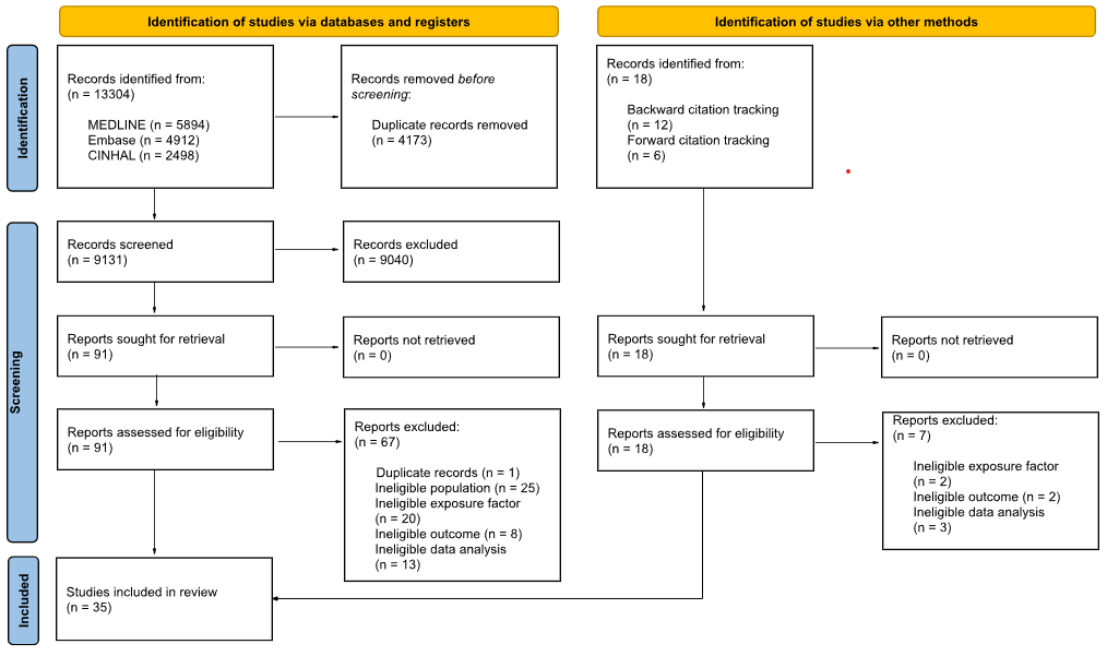
    From 9 054 initial records, 24 studies met the inclusion criteria, all being case reports. The most frequently reported red flags were severe neck pain, which may become progressive and constant, neurological signs and symptoms, and nocturnal neck pain. However, inconsistencies in reporting were observed: many commonly discussed red flags were absent from case reports, while some red flags highlighted in case reports are not extensively covered in the literature.

    Conclusion

    Evidence on red flags for tumours in patients with neck pain in primary care remains fragmented. While certain red flags were frequently reported, their clinical utility is limited by inconsistent reporting and a lack of robust evidence. Further research is needed to standardize red flags and assess their diagnostic accuracy to improve early tumour detection in patients with neck pain.

    REFERENCES

    1. Mourad F, Giovannico G, Maselli F, Bonetti F, Fernández de las Peñas C, Dunning J. Basilar impression presenting as intermittent mechanical neck pain: a rare case report. BMC Musculoskelet Disord. 11 gennaio 2016; 17:7.
    2. Safiri S, Kolahi AA, Hoy D, Buchbinder R, Mansournia MA, Bettampadi D, et al. Global, regional, and national burden of neck pain in the general population, 1990-2017: systematic analysis of the Global Burden of Disease Study 2017. BMJ. 26 marzo 2020;368:m791.
    3. Vos T, Lim SS, Abbafati C, Abbas KM, Abbasi M, Abbasifard M, et al. Global burden of 369 diseases and injuries in 204 countries and territories, 1990–2019: a systematic analysis for the Global Burden of Disease Study 2019. The Lancet. 17 ottobre 2020;396(10258):1204–22.
    4. GBD 2021 Neck Pain Collaborators. Global, regional, and national burden of neck pain, 1990-2020, and projections to 2050: a systematic analysis of the Global Burden of Disease Study 2021. Lancet Rheumatol. marzo 2024;6(3):e142–55.
    5. Masters S. Acute cervical spine pain in primary care. Aust J Gen Pract. 1 novembre 2023;52(11):745–50.
    6. Mourad F, Cataldi F, Patuzzo A, Tunnera S, Dunning J, Fernández-de-las-Peñas C, et al. Craniopharyngioma in a young woman with symptoms presenting as mechanical neck pain associated with cervicogenic headache: a case report. Physiother Theory Pract. 2021;37(4):549–58.
    7. Finucane LM, Downie A, Mercer C, Greenhalgh SM, Boissonnault WG, Pool-Goudzwaard AL, et al. International Framework for Red Flags for Potential Serious Spinal Pathologies. J Orthop Sports Phys Ther. luglio 2020;50(7):350–72.
    8. Feller D, Chiarotto A, Koes B, Maselli F, Mourad F. Red flags for potential serious pathologies in people with neck pain: a systematic review of clinical practice guidelines. Arch Physiother. 2024;14:105–15.
    9. Maselli F, Palladino M, Barbari V, Storari L, Rossettini G, Testa M. The diagnostic value of Red Flags in thoracolumbar pain: a systematic review. Disabil Rehabil. aprile 2022;44(8):1190–206.
  • MODELLI DI PREDIZIONE DIAGNOSTICA PER LE FRATTURE VERTEBRALI IN PERSONE CON DOLORE SPINALE O TRAUMA: UNA REVISIONE SISTEMATICA CON META-ANALISI

    MODELLI DI PREDIZIONE DIAGNOSTICA PER LE FRATTURE VERTEBRALI IN PERSONE CON DOLORE SPINALE O TRAUMA: UNA REVISIONE SISTEMATICA CON META-ANALISI

    DIAGNOSTIC PREDICTION MODELS FOR SPINAL FRACTURES IN PEOPLE WITH SPINAL PAIN OR TRAUMA: A SYSTEMATIC REVIEW WITH META-ANALYSIS

    Autori

    Feller Daniel (Azienda Provinciale per i Servizi Sanitari, Trento, Italia; Erasmus Medical Center, Rotterdam, The Netherlands)

    Wingbermühle Roel (Erasmus Medical Center, Rotterdam, The Netherlands)

    Oei Edwin (Erasmus Medical Center, Rotterdam, The Netherlands)

    Koes Bart (Erasmus Medical Center, Rotterdam, The Netherlands; University of Southern Denmark, Odense, Denmark)

    Chiarotto Alessandro (Erasmus Medical Center, Rotterdam, The Netherlands)

    Background and aims

    In this systematic review we aimed to evaluate the performance of multivariable diagnostic prediction models for identifying spinal fractures in patients with spinal pain and/or trauma.

    Methods

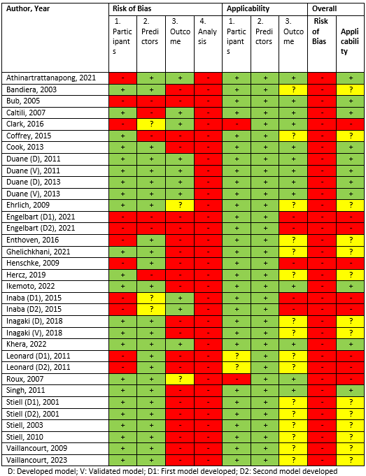
    We conducted a systematic review with meta-analysis, following a prospectively registered protocol in PROSPERO (CRD42024539898). We included studies that developed (with or without internal validation) or externally validated multivariable diagnostic prediction models for identifying spinal fractures in patients with spinal pain and/or trauma. We searched MEDLINE, EMBASE, and Web of Science (up to May 2025), and conducted backward and forward citation tracking strategies. Two independent reviewers extracted studies’ data using the CHARMS checklist, and assessed the risk of bias with the PROBAST tool. A bivariate random-effects model was used to pool classification measures, while univariate random-effects models were used for discrimination and calibration measures. We assessed the certainty of the evidence using an adapted version of the GRADE for prognostic factor studies.

    Results

    We included 27 studies encompassing 34 diagnostic models. All models showed an overall high risk of bias. Meta-analyses of ten studies that externally validated the use of the Canadian C-spine Rule in adults presenting with trauma to emergency departments demonstrated, with very low certainty of the evidence, excellent sensitivity (0.999; 95% CI 0.976 to 1), an high area under the curve (0.85; 95% CI 0.72 to 0.97), and a low specificity (0.188; 95% CI 0.063 to 0.443). We estimated a pooled positive likelihood ratio of 1.23 (95% CI 0.978 to 1.548) and a negative likelihood ratio of 0.007 (95% CI 0.001 to 0.082) for the same model. Other models for traumatic cervical fractures and osteoporotic fractures showed promise but lacked external validation or sufficient reporting on calibration and discrimination measures. No models for thoracolumbar fractures were deemed ready to be used clinically.

    Conclusion

    Our findings indicate that, while the Canadian C-spine Rule shows potential for screening traumatic cervical fractures, the very low to low certainty of the evidence limits confidence in its accuracy and appropriateness for clinical use. Notably, the Canadian C-spine Rule has only been validated in emergency department settings and is therefore not applicable to standard rehabilitation settings. Also, we did not identify any externally validated model suitable for clinical use regarding osteoporotic fractures, traumatic fractures of the thoracolumbar spine, and traumatic fractures of the cervical spine in non-emergency settings. Future research with rigorous methodological and statistical approaches should aim to fill these gaps.

    REFERENCES

    1. Moons KG, de Groot JA, Bouwmeester W, Vergouwe Y, Mallett S, Altman DG, Reitsma JB, Collins GS. Critical appraisal and data extraction for systematic reviews of prediction modelling studies: the CHARMS checklist. PLoS Med. 2014 Oct 14;11(10):e1001744. doi: 10.1371/journal.pmed.1001744. PMID: 25314315; PMCID: PMC4196729.
    2. Wolff RF, Moons KGM, Riley RD, Whiting PF, Westwood M, Collins GS, Reitsma JB, Kleijnen J, Mallett S; PROBAST Group†. PROBAST: A Tool to Assess the Risk of Bias and Applicability of Prediction Model Studies. Ann Intern Med. 2019 Jan 1;170(1):51-58. doi: 10.7326/M18-1376. PMID: 30596875.
    3. Huguet A, Hayden JA, Stinson J, McGrath PJ, Chambers CT, Tougas ME, Wozney L. Judging the quality of evidence in reviews of prognostic factor research: adapting the GRADE framework. Syst Rev. 2013 Sep 5;2:71. doi: 10.1186/2046-4053-2-71. PMID: 24007720; PMCID: PMC3930077.
    4. van Smeden M, Reitsma JB, Riley RD, Collins GS, Moons KG. Clinical prediction models: diagnosis versus prognosis. J Clin Epidemiol. 2021 Apr;132:142-145. doi: 10.1016/j.jclinepi.2021.01.009. PMID: 33775387.
    5. Feller D, Chiarotto A, Koes B, Maselli F, Mourad F. Red flags for potential serious pathologies in people with neck pain: a systematic review of clinical practice guidelines. Arch Physiother. 2024 Dec 4;14:105-115. doi: 10.33393/aop.2024.3245. PMID: 39639931; PMCID: PMC11618059.
  • IL RANGE OF MOTION COME FATTORE PROGNOSTICO NEI PAZIENTI CON DOLORE SPINALE SPECIFICO E NON SPECIFICO: UNA REVISIONE SISTEMATICA

    IL RANGE OF MOTION COME FATTORE PROGNOSTICO NEI PAZIENTI CON DOLORE SPINALE SPECIFICO E NON SPECIFICO: UNA REVISIONE SISTEMATICA

    RANGE OF MOTION AS A PROGNOSTIC FACTOR IN PATIENTS WITH SPECIFIC AND NON–SPECIFIC SPINAL PAIN: A SYSTEMATIC REVIEW

    Autori

    Feller Daniel (Azienda Provinciale per i Servizi Sanitari, Trento, Italia; Erasmus Medical Center, Rotterdam, The Netherlands)

    Bortoli Michela (Azienda Provinciale per i Servizi Sanitari, Trento, Italia)

    Rigo Adriano (Azienda Provinciale per i Servizi Sanitari, Trento, Italia)

    Maini Irene (Azienda Provinciale per i Servizi Sanitari, Trento, Italia)

    Hayden Jill (Dalhousie University, Halifax, Canada)

    Chiarotto Alessandro (Erasmus Medical Center, Rotterdam, The Netherlands)

    Background and aims

    Identifying prognostic factors—variables that help predict clinical outcomes—is essential for improving patient care and guiding treatment decisions. Range of motion (ROM) is routinely assessed in clinical practice, yet its prognostic value in patients with spinal pain remains unclear. Therefore, this systematic review aimed to determine whether reduced spinal ROM predicts outcomes such as pain intensity, disability, or global recovery in individuals with specific or non-specific spinal pain.

    Methods

    (CRD42024583518). We included prospective and retrospective cohort studies, case-control studies, and secondary analyses of randomized controlled trials evaluating ROM as a prognostic factor in adults (≥18 years) with spinal pain. We included studies assessing active or passive ROM using any objective method. Databases searched included MEDLINE, Embase, CENTRAL, and CINAHL up to May 24, 2024. Risk of bias was assessed using the QUIPS tool, and certainty of evidence was rated using the GRADE approach. Two authors independetly performed the study selection, data extraction and risk of bias assessment phases. Due to clinical and methodological heterogeneity, results were synthesized narratively.

    Results

    A total of 35 studies were included: 11 on acute low back pain (LBP), 10 on chronic or unspecified LBP, 4 on chronic neck pain, 2 on lumbar spinal stenosis, and 8 on whiplash-associated disorders. No studies addressed acute neck pain. ROM was most commonly measured using inclinometers and goniometers, though methods varied widely. Across all conditions, the certainty of evidence was very low due to high risk of bias (particularly inadequate adjustment for confounders), inconsistent findings, and methodological heterogeneity. While some studies reported significant associations between reduced ROM and worse outcomes (especially in whiplash and lumbar stenosis), most showed no consistent relationship. Many studies showed inadequate reporting, often presenting only statistical test results without point estimates or confidence intervals, which limits the clinical interpretability of the findings.

    Conclusion

    There is currently low to very low-certainty evidence that reduced spinal ROM is a prognostic factor for pain, disability, or recovery in patients with spinal pain. Therefore, routine ROM assessment may have limited value for prognosis alone. However, ROM remains clinically relevant for identifying movement-related dysfunction and informing treatment decisions. Future high-quality, prospective studies with robust adjustment for confounders are needed to clarify the independent prognostic role of spinal ROM, especially in underexplored populations such as those with acute neck pain.

    REFERENCES

    1. Hayden JA, van der Windt DA, Cartwright JL, Côté P, Bombardier C. Assessing bias in studies of prognostic factors. Ann Intern Med. 2013 Feb 19;158(4):280-6. doi: 10.7326/0003-4819-158-4-201302190-00009. PMID: 23420236.
    2. Huguet A, Hayden JA, Stinson J, McGrath PJ, Chambers CT, Tougas ME, Wozney L. Judging the quality of evidence in reviews of prognostic factor research: adapting the GRADE framework. Syst Rev. 2013 Sep 5;2:71. doi: 10.1186/2046-4053-2-71. PMID: 24007720; PMCID: PMC3930077.
    3. Riley RD, Hayden JA, Steyerberg EW, Moons KG, Abrams K, Kyzas PA, Malats N, Briggs A, Schroter S, Altman DG, Hemingway H; PROGRESS Group. Prognosis Research Strategy (PROGRESS) 2: prognostic factor research. PLoS Med. 2013;10(2):e1001380. doi: 10.1371/journal.pmed.1001380. Epub 2013 Feb 5. PMID: 23393429; PMCID: PMC3564757.
  • Performance delle referral strategies per pazienti con sospetta spondiloartrite assiale: una revisione sistematica

    Performance of referral strategies for patients with suspected axial spondyloarthritis: a systematic review

    Autori

    Feller Daniel (Azienda Provinciale per i Servizi Sanitari, Trento, Italia; Erasmus Medical Center, Rotterdam, The Netherlands)

    Wingbermühle Roel (Erasmus Medical Center, Rotterdam, The Netherlands)

    Koes Bart (Erasmus Medical Center, Rotterdam, The Netherlands)

    Ramiro Sofia (Leiden University Medical Center, Leiden, The Netherlands)

    Chiarotto Alessandro (Erasmus Medical Center, Rotterdam, The Netherlands)

    Background and aims

    Axial spondyloarthritis (axSpA) is a chronic inflammatory condition often misdiagnosed as non-specific low back pain, leading to diagnostic delays averaging 6.7 years. Early referral to rheumatology is essential to initiate timely treatment and prevent structural damage. Various referral strategies have been proposed to assist non-specialists in identifying patients with suspected axSpA. However, the diagnostic performance of these strategies has not been comprehensively assessed since the 2012 ASAS review, which lacked methodological rigor. This systematic review aims to evaluate the diagnostic accuracy of referral strategies—defined as diagnostic tests or models—for identifying patients suspected of having axSpA.

    Methods

    We conducted a systematic review following a protocol registered on PROSPERO (CRD42025634153). We systematically searched MEDLINE, Embase, and CINAHL, with additional backward and forward citation tracking using Web of Science. No restrictions were placed on language or publication year. Two reviewers independently screened titles, abstracts, and full texts using Covidence, resolving disagreements via consensus or a third reviewer. Eligible studies included cross-sectional, cohort, and case-control designs that assessed referral strategies in adults with chronic (>3 months) low back pain. Data extraction will follow the CHARMS checklist for diagnostic models and included sensitivity, specificity, likelihood ratios, and AUC. Risk of bias will be assessed using QUADAS-2 (for diagnostic tests) and PROBAST (for diagnostic models). Meta-analysis or narrative synthesis will be conducted depending on heterogeneity, with evidence quality assessed using GRADE-based approaches adapted for diagnostic accuracy and prediction models.

    Results

    Following duplicate removal and screening, we included 21 studies evaluating the diagnostic performance of referral strategies for axSpA. These studies span various healthcare settings and geographic regions, and assess both test-based criteria (e.g., Berlin criteria) and multivariable prediction models. Data extraction and risk-of-bias assessment are currently ongoing. The final analysis, including meta-analyses and subgroup assessments, will be completed by August 2025.

    Conclusion

    This systematic review will provide updated evidence on the diagnostic accuracy of referral strategies for axSpA, supporting clinicians and policymakers in choosing appropriate tools for early identification. Findings aim to reduce diagnostic delays and improve patient outcomes through optimized referral practices.

    REFERENCES

    1. Navarro-Compán V, Sepriano A, El-Zorkany B, van der Heijde D. Axial spondyloarthritis. Ann Rheum Dis. 2021 Dec;80(12):1511-1521. doi: 10.1136/annrheumdis-2021-221035. Epub 2021 Oct 6. PMID: 34615639.
    2. Zhao SS, Pittam B, Harrison NL, Ahmed AE, Goodson NJ, Hughes DM. Diagnostic delay in axial spondyloarthritis: a systematic review and meta-analysis. Rheumatology (Oxford). 2021 Apr 6;60(4):1620-1628. doi: 10.1093/rheumatology/keaa807. PMID: 33428758.
    3. Whiting PF, Rutjes AW, Westwood ME, Mallett S, Deeks JJ, Reitsma JB, Leeflang MM, Sterne JA, Bossuyt PM; QUADAS-2 Group. QUADAS-2: a revised tool for the quality assessment of diagnostic accuracy studies. Ann Intern Med. 2011 Oct 18;155(8):529-36. doi: 10.7326/0003-4819-155-8-201110180-00009. PMID: 22007046.
    4. Wolff RF, Moons KGM, Riley RD, Whiting PF, Westwood M, Collins GS, Reitsma JB, Kleijnen J, Mallett S; PROBAST Group†. PROBAST: A Tool to Assess the Risk of Bias and Applicability of Prediction Model Studies. Ann Intern Med. 2019 Jan 1;170(1):51-58. doi: 10.7326/M18-1376. PMID: 30596875.
  • Dolore alla spalla: Immagini diagnostiche sì o no?

    Dolore alla spalla: Immagini diagnostiche sì o no?

    Shoulder pain: To image or not to image?

    Autori

    Brindisino Fabrizio (Department of Medicine and Health Science “Vincenzo Tiberio”, University of Molise, Campobasso, Italy)

    Paul Salamh (Krannert School of Physical Therapy, University of Indianapolis, 1400 East Hanna Avenue, Indianapolis, IN)

    Chad Cook (Department of Population Health Sciences, Duke University, Durham, NC, USA)

    Jeremy Lewis (Therapy Department, Central London Community Healthcare National Health Service Trust, Finchley Memorial Hospital, London, UK)

    Alvisa Palese (Department of Medical Sciences, University of Udine, Udine, Italy)

    Germano Guerra (Department of Medicine and Health Science “Vincenzo Tiberio”, University of Molise, Campobasso, Italy)

    Jacopo Bonavita (Neurorehabilitation Unit, Villa Rosa Hospital, APSS Trento, Trento, Italy)

    Giacomo Rossettini (School of Physiotherapy, University of Verona, Verona, Italy)

    Background and aims

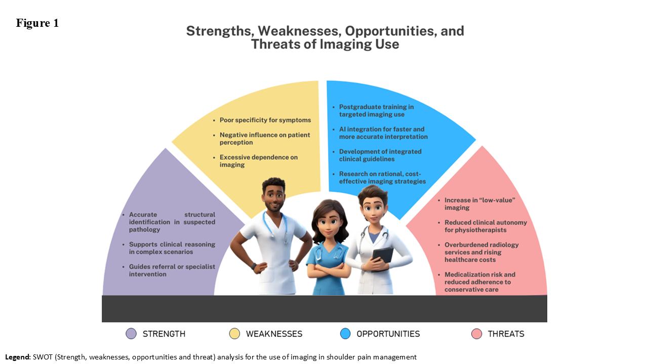
    Imaging findings should be interpreted within the broader context of an individual’s shoulder symptoms [1]. While imaging is valuable in identifying specific structural pathologies, findings are often equivocal. Defining what imaging changes constitute “normal”, “abnormal”, “unrelated”, “solely causative” findings, and which are, “contributory”, or “associated” with symptoms is a clinical minefield. It is arguable that many so called “abnormalities” have been labeled as such because they represent deviations in structure from idealized and flawless anatomical drawings.

    Methods

    To promote consideration for the role of “bio” in shoulder symptoms and encourage debate among clinicians and researchers, this opinion paper aims to stimulate discussion on the value of imaging in management of musculoskeletal shoulder pain. We sought to capture diverse perspectives by incorporating input from various health disciplines working within the musculoskeletal field, aimed at providing a comprehensive perspective to our manuscript.

    Results

    Although the judicious requests of imaging may improve clinician understanding of a specific clinical scenario, between 20–50% of imaging requests are inappropriate or of “low value” (e.g., a service offering no or minimal benefit) [2]. Conversely, strategies aimed at reducing low-value imaging have the potential to decrease costs by as much as 95% without compromising patient well-being [2].

    Studies investigating this issue, question the clinical utility of routine imaging for treatment planning, given the high prevalence of anatomical variations in asymptomatic shoulders [1,3]. Indeed, while imaging may identify tissue pathology, it often cannot reliably determine the clinical significance of these findings or their correlation with specific symptoms. Acceptance of imaging findings (i) without defining what abnormal is based upon and (ii) without stating the prevalence of such findings in people without symptoms, may lead to over medicalization and detrimentally impact on healthcare sustainability [4].

    Conclusion

    Clinical practice frequently lacks balance and frequently is conducted at the ends of a spectrum. With one end being rigid reliance on structural imaging and the other its rejection in favor of a psychosocial narrative. To promote a judicious, resourceful, and evidence-based utilization of imaging, research should emphasize a comprehensive bio-psycho-social care pathway. Within this framework, all relevant domains should be engaged and weighted according to the specific clinical context. A balanced, context-driven use of imaging, grounded in clinical reasoning and embedded in a biopsychosocial model, can enhance diagnosis, inform treatment, and support shared decision-making. Imaging is a valuable tool when used wisely: the question is not whether to use it, but when, why, and for whom.

    REFERENCES

    [1] Tran G, Cowling P, Smith T, Bury J, Lucas A, Barr A, et al. What Imaging-Detected Pathologies Are Associated With Shoulder Symptoms and Their Persistence? A Systematic Literature Review. Arthritis Care & Research. 2018;70:1169-84.

    [2] Kjelle E, Brandsæter IØ, Andersen ER, Hofmann BM. Cost of Low-Value Imaging Worldwide: A Systematic Review. Applied Health Economics and Health Policy. 2024;22:485-501.

    [3] Gill TK, Shanahan EM, Allison D, Alcorn D, Hill CL. Prevalence of abnormalities on shoulder MRI in symptomatic and asymptomatic older adults. Int J Rheum Dis. 2014;17:863-71.

    [4] Ingraham B, Miller K, Iaia A, Sneider MB, Naqvi S, Evans K, et al. Reductions in High-End Imaging Utilization With Radiology Review and Consultation. Journal of the American College of Radiology : JACR. 2016;13:1079-82

  • Le informazioni fornite dai sanitari sono associate a prospettiva di recupero, emozioni e catastrofismo in soggetti con spalla congelata. Uno studio Trasversale

    Healthcare professional information impacts frozen shoulder subjects’ recovery outlook, emotions, and catastrophizing. A cross-sectional study.

    Autori

    Brindisino Fabrizio (Department of Medicine and Health Science “Vincenzo Tiberio”, University of Molise, Campobasso, Italy)

    Rossettini Giacomo (School of Physiotherapy, University of Verona, Verona, Italy)

    Andrea De Santis (Department of Medicine and Health Science “Vincenzo Tiberio”, University of Molise, Campobasso, Italy)

    Crestani Mauro (Department of Medicine and Health Science “Vincenzo Tiberio”, University of Molise, Campobasso, Italy)

    Turolla Andrea (Department of Biomedical and Neuromotor Sciences, Alma Mater Studiorum University of Bologna, Italy)

    Background and aims

    The significance of patient-clinician communication is acknowledged within broader musculoskeletal contexts [1,2]. However, the specific impact of information provided during the initial physiotherapy consultation on recovery expectations, emotional state, and catastrophizing thoughts in patients with frozen shoulder (FS) has not been previously investigated. This study aims to examine the influence of clinician-communicated information on recovery expectations, emotional state, and catastrophizing thoughts in individuals with FS.

    Methods

    This cross-sectional exploratory online survey followed the Strengthening the Reporting of Observational Studies in Epidemiology (STROBE) guidelines [3]. Participants clinically diagnosed with FS according to Kelley’s criteria [4] completed a 32-item closed-multiple choice online questionnaire via Google Forms. Subjects were recruited through physiotherapy practices in Italy, between April and July 2023.

    Results

    A total of 110 Italian subjects diagnosed with FS were prospectively recruited for this study. Most participants reported feeling adequately informed by helpful, reassuring, and encouraging explanations, and they expressed a significant need for reassurance. Recovery expectations were generally high, while their emotional state was reported as lower. The most prevalent catastrophizing thoughts reported included the belief that FS is a long-term condition, that the pain is severe and persistent, and that they would never be able to return to previous activities. Participants also frequently reported feeling overwhelmed by FS.

    Respondents who received unhelpful explanations —those that did not improve their ability to manage FS, increased anxiety and concerns about potential recovery failure, lacked reassurance, did not help manage pessimistic thoughts about FS, or were discouraging (e.g., increasing fear of movement)— were predominantly associated with negative recovery expectations. Conversely, a negative emotional state did not appear to be associated with perceived worrying, unhelpful, or un-reassuring information. Interestingly, some catastrophizing thoughts (e.g., “I will never raise my arm as I used to,” “the pain was never-ending,” “all efforts to heal were useless,” “I felt overwhelmed by FS,” “being afraid that FS is a long-term pathology”) did not seem to be associated with perceived worrying, unhelpful, or un-reassuring information, while others were.

    Conclusion

    The content and style of clinician communication play a crucial role in shaping recovery expectations among FS patients, potentially impacting treatment outcomes. It appears that emotional state might be influenced by variables other than perceived information. Lastly, catastrophizing thoughts should be mitigated through helpful, encouraging, and reassuring communication. Employing advanced communication skills and tailoring information delivery could increase recovery expectation and reduce catastrophic thinking, thereby fostering improved adherence and engagement in physiotherapy.

    REFERENCES

    [1] Rossettini G, Palese A, Geri T, Fiorio M, Colloca L, TestaM (2018) Physical therapists’ perspectives on using contextual factors in clinical practice: Findings from an Italian national survey. PLoS ONE 13(11): e0208159. https://doi.org/10.1371/journal.pone.0208159

    [2] Palese A, Cadorin L, Testa M, Geri T, Colloca L, Rossettini G. Contextual factors triggering placebo and nocebo effects in nursing practice: Findings from a national cross-sectional study. J Clin Nurs. 2019 May;28(9-10):1966-1978. doi: 10.1111/jocn.14809. Epub 2019 Feb 18. PMID: 30706543.

    [3] von Elm E, Altman DG, Egger M, Pocock SJ, Gøtzsche PC, Vandenbroucke JP. The Strengthening the Reporting of Observational Studies in Epidemiology (STROBE) Statement: guidelines for reporting observational studies. International journal of surgery (London, England). 2014;12:1495-9.

    [4] Kelley MJ, Shaffer MA, Kuhn JE, Michener LA, Seitz AL, Uhl TL, et al. Shoulder pain and mobility deficits: adhesive capsulitis. The Journal of orthopaedic and sports physical therapy. 2013;43:A1-31

  • Associazini tra intensità di dolore, range di movimento, disabilità e qualità della vita correlata allo stato di salute in soggetti con frozen shoulder. Uno studio trasversale

    Associazini tra intensità di dolore, range di movimento, disabilità e qualità della vita correlata allo stato di salute in soggetti con frozen shoulder. Uno studio trasversale

    Association Between Pain Intensity, Range of Motion, Disability, and Health-Related Quality of Life Variables in Frozen Shoulder: A Cross-Sectional Study

    Autori

    Brindisino Fabrizio (Department of Medicine and Health Science “Vincenzo Tiberio”, University of Molise, Campobasso, Italy)

    Feller Daniel (Department of General Practice, Erasmus MC, University Medical Center, Rotterdam, The Netherlands)

    Andriesse Arianna (Medical Translation Private Practice c/o Andriesse Medical Translator, Lecce, Italy)

    Venturin Davide (Department of Medicine and Health Science “Vincenzo Tiberio”, University of Molise, Campobasso, Italy)

    Poser Antonio (Physiotherapy private practice Kinè c/o Viale Venezia 13/Q San Vendemiano, Italy)

    Background and aims

    While the relationship between disability and pain is well established [1], recent studies have highlighted the association between health-related, social and psychological factors, and negative functional outcomes in patients with Frozen Shoulder (FS) [2]. This study analyzes the association between pain, disability, health-related quality of life domains, and range of motion (ROM) during the first physiotherapy consultation in FS patients.

    Methods

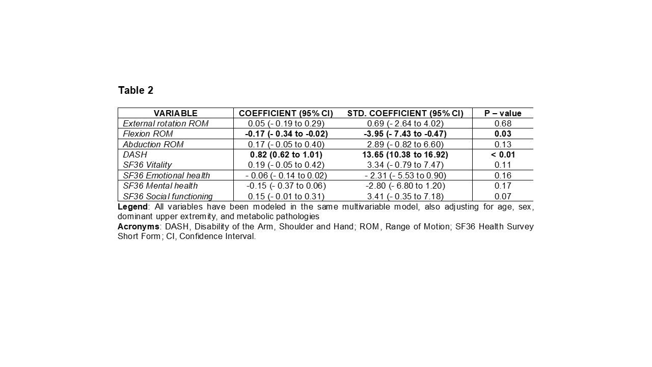
    This cross-sectional study followed the STROBE guidelines [3]. Participants were recruited between January and October 2024. Each participant anonymously filled out the Shoulder Pain and Disability Index, the Disabilities of the Arm, Shoulder, and Hand (DASH) questionnaire, and the 36-Item Short Form Health Survey. ROM restrictions in flexion, abduction, and external rotation with the arm by the side were assessed. Statistical analysis was performed using multivariable linear regression —modeling all independent variables simultaneously— and adjusting for confounding factors.

    Results

    A total of 149 subjects were included (53% female). Significant correlations were found between pain and disability (standardized coefficient = 13.65, 95% CI 10.38 to 16.92, p < 0.01), as well as between pain and flexion restriction (standardized coefficient = -3.95 95%CI -7.43 to -0.47, p=0.03).

    Conclusion

    Our findings confirm that pain is a primary determinant of disability in FS patients, even at early stages. A significant association between pain and flexion limitation was also observed, with flexion being the most sensitive movement to pain. The lack of correlation with health-related quality of life variables suggests that, while pain limits physical function, its psychological impact—assessed through such outcome measures used—may not yet be evident. This study provides strong evidence of the association among pain, disability, and ROM in flexion during the initial physiotherapy consultation for FS, suggesting that early clinical focus on pain management may help reduce disability, improving overall patient outcomes.

    REFERENCES

    [1] Mertens MG, Struyf F, Verborgt O, Dueñas L, Balasch-Bernat M, Navarro-Ledesma S, et al. Exploration of the clinical course and longitudinal correlations in frozen shoulder: The role of autonomic function, central pain processing, and psychological variables. A longitudinal multicenter prospective observational study. Musculoskeletal Science and Practice. 2023;67:102857.

    [2] Brindisino F, Silvestri E, Gallo C, Venturin D, Di Giacomo G, Peebles AM, et al. Depression and Anxiety Are Associated With Worse Subjective and Functional Baseline Scores in Patients With Frozen Shoulder Contracture Syndrome: A Systematic Review. Arthroscopy, sports medicine, and rehabilitation. 2022;4:e1219-e34.

    [3] von Elm E, Altman DG, Egger M, Pocock SJ, Gøtzsche PC, Vandenbroucke JP. The Strengthening the Reporting of Observational Studies in Epidemiology (STROBE) Statement: guidelines for reporting observational studies. International journal of surgery (London, England). 2014;12:1495-9


  • Advancing from Item-Level to Total Score-Based diagnosis in the Coma Recovery Scale–Revised: Evidence from a Diagnostic Accuracy Study

    Advancing from Item-Level to Total Score-Based diagnosis in the Coma Recovery Scale–Revised: Evidence from a Diagnostic Accuracy Study

    Passaggio dalla diagnosi basata sugli item a quella sul punteggio totale nella Coma Recovery Scale–Revised: uno studio di accuratezza diagnostica

    Autori

    Caselli Serena (Unità Operativa Complessa di Medicina Riabilitativa, Azienda Ospedaliero-Universitaria di Modena, Modena, Italy)

    Pellicciari Leonardo (IRCCS Istituto delle Scienze Neurologiche di Bologna, Bologna, Italy)

    Leonardi Matilde (SC Neurologia, Salute Pubblica, Disabilità, Fondazione IRCCS Istituto Neurologico Carlo Besta, Milan, Italy)

    Magnani Francesca Giulia (SC Neurologia, Salute Pubblica, Disabilità, Fondazione IRCCS Istituto Neurologico Carlo Besta, Milan, Italy)

    Cacciatore Martina (SC Neurologia, Salute Pubblica, Disabilità, Fondazione IRCCS Istituto Neurologico Carlo Besta, Milan, Italy)

    Barbadoro Filippo (SC Neurologia, Salute Pubblica, Disabilità, Fondazione IRCCS Istituto Neurologico Carlo Besta, Milan, Italy)

    Ippoliti Camilla (SC Neurologia, Salute Pubblica, Disabilità, Fondazione IRCCS Istituto Neurologico Carlo Besta, Milan, Italy)

    Kreiner Svend (Section of Biostatistics, Department of Public Health, University of Copenhagen, Copenhagen, Denmark)

    La Porta Fabio (IRCCS Istituto delle Scienze Neurologiche di Bologna, Bologna, Italy)

    Background and aims

    The Coma Recovery Scale-Revised (CRS-R) is the gold standard for diagnosing patients with Disorder of Consciousness (DOC). Five items out of six provide scores linked to a diagnosis of Unresponsive Wakefulness Syndrome (UWS), Minimally Conscious State (MCS), or emergence from MCS (eMCS). However, no diagnostic criteria are linked to the total score.

    Therefore, this study proposes to define and compare the diagnostic accuracy of total score cutoffs based on item-level diagnostic criteria proposed recently[1] as a reference standard, and to improve the diagnostic accuracy of these total score cutoffs using a modified version of the original rule (i.e., the diagnostic category coincides with the highest category defined at least by one item)[2] currently in use.

    Methods

    Patients with DOC as a consequence of a severe acquired brain injury and admitted to post-acute and follow-up neuro-rehabilitation services were included. Diagnostic accuracy (DA) of the total score cutoffs based on each of the item-level diagnostic criteria set proposed recently[1-4], and the agreement indexes between item-level and total score-level diagnosis were computed. Finally, the comparison of the diagnostic accuracy of the total score cutoffs according to Giacino’s original rule[2] and the “modified rule” (i.e., the diagnostic category coincides with the highest category defined at least by two/three/four items) with Caselli’s criteria[1] was performed.

    Results

    380 patients with DOC (mean±SD: 52.1±16.8 years, 65% male) were included for a total of 727 CRS-R assessments.

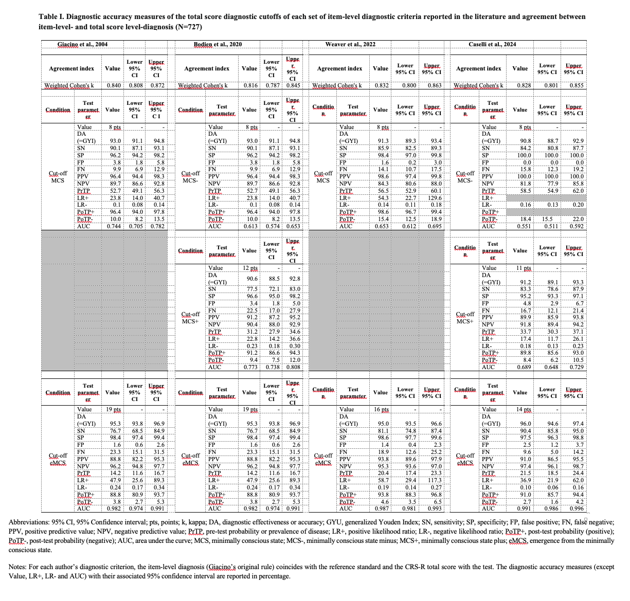
    The comparison of total score cutoffs for each diagnostic criterion showed that for MCSplus and eMCS, the DA value is 90-95% (Table I), with high false negatives (FN) for most criteria sets (MCSplus: Bodien 12pt 22.5%; Caselli 11pt 16.7%; eMCS: Giacino and Bodien 19pt 23%; Weaver 16pt 19%). For MCSminus, the four cutoffs (8pt) had high specificity, essential for identifying patients with early signs of consciousness. Weighted Cohen’s k between the item and total score diagnoses was 0.82 for all four criteria sets.

    Applying the “modified rule” with Caselli’s criteria resulted in the highest DA values for both MCSplus (14pt 97%) and eMCS (19pt 99%), with a lower FN rate (<11%) (Tables II and III).

    Conclusion

    Being the diagnosis based on individual items, the initial total score cutoffs partly addressed the low reliability of item-level cutoffs. The introduction of the “modified rule,” which requires at least two items per diagnostic category for diagnosis, allowed for advancing from item-level to a reliable total score-based diagnosis with less measurement error.

    REFERENCES

    1. Caselli S, Leonardi M, Magnani FG, Cacciatore M, Barbadoro F, Ippoliti C, Kreiner S, Pellicciari L, La Porta F. Comparing the Different Sets of Item-Level Diagnostic Criteria of the Coma Recovery Scale-Revised (CRS-R): A Measurement-Based Approach Driven by Rasch Analysis. Arch Phys Med Rehabil. 2024 Dec 18:S0003-9993(24)01406-0. doi: 10.1016/j.apmr.2024.12.009.
    2. Giacino JT, Kalmar K, Whyte J. The JFK Coma Recovery Scale-Revised: measurement characteristics and diagnostic utility. Arch Phys Med Rehabil. 2004 Dec;85(12):2020-9. doi: 10.1016/j.apmr.2004.02.033.
    3. Bodien YG, Chatelle C, Taubert A, Uchani S, Giacino J T, Ehrlich-Jones L. Updated measurement characteristics and clinical utility of the Coma Recovery Scale-Revised among individuals with acquired brain injury. Arch Phys Med Rehabil. 2021;102(1):169-171. doi: 10.1016/j.apmr.2020.09.369.
    4. Weaver JA, Cogan AM, O’Brien KA, Hansen P, Giacino JT, Whyte J, Bender Pape T, van der Wees P, Mallinson T. Determining the Hierarchy of Coma Recovery Scale-Revised Rating Scale Categories and Alignment with Aspen Consensus Criteria for Patients with Brain Injury: A Rasch Analysis. J Neurotrauma. 2022 Oct;39(19-20):1417-1428. doi: 10.1089/neu.2022.0095.
  • La Funzione del riflesso vestibolo-oculomotore è alterata in persone con Malattia di Parkinson: uno studio trasversale con il video Head Impulse Test

    Vestibulo-oculomotor reflex function is altered in people with Parkinson’s Disease: a cross-sectional study using the video Head Impulse Test

    Autori

    Diego Piatti (Santa Lucia Foundation, Scientific Institute for Research and Health Care, 00179 Rome, Italy)

    Laura Casagrande Conti (Santa Lucia Foundation, Scientific Institute for Research and Health Care, 00179 Rome, Italy)

    Leonardo Manzari (MSA ENT Academy Center, 03043 Cassino, Italy)

    Domenico De Angelis (Santa Lucia Foundation, Scientific Institute for Research and Health Care, 00179 Rome, Italy)

    Ugo Nocentini (Santa Lucia Foundation, Scientific Institute for Research and Health Care, 00179 Rome, Italy)

    Marco Tramontano (Department of Biomedical and Neuromotor Sciences, University of Bologna, 40138 Bologna, Italy; Unit of Occupational Medicine, IRCCS Azienda Ospedaliero-Universitaria di Bologna, 40138 Bologna, Italy)

    Nicola Ferri (Department of Biomedical and Neuromotor Sciences, University of Bologna, 40138 Bologna, Italy)

    Background and aims

    Parkinson’s Disease (PD) is a neurodegenerative, progressive disorder known for motor and non-motor symptoms (1). Vestibular dysfunction in PD may exacerbate postural instability and gait disturbances. The vestibular system, via the vestibulo-ocular reflex (VOR), is crucial for maintaining dynamic gaze stability, and its role in PD is raising interest among researchers (2). However, the prevalence of vestibular dysfunctions remains unclear how prevalent vestibular dysfunctions are isn’t fully clear. This study aims to objectively investigate the VOR function in people with PD using the video Head Impulse Test.

    Methods

    This is a cross-sectional study conducted in a neurorehabilitation hospital. People with PD were included if they had no dementia and the ability to walk without physical assistance. The video Head Impulse Test was used to assess the VOR function in the six semicircular canals. Both the Head Impulse Paradigm (HIMP) and the Suppression Head Impulse Paradigm (SHIMP) paradigms were considered.

    Results

    Thirty-five people with PD (mean age: 69.9±8.4; 11 females) with moderate motor symptoms (UPDRS-part III: 27.7 ± 6.8) were included. Using normative cut-offs, 69% of the participants had at least one dysfunctional canal (60% hypo-gain, 9% hyper-gain). The prevalence reached 83% when both the HIMP and SHIMP paradigms were considered.

    Conclusion

    There is a high prevalence of vestibular dysfunction in people with PD. The instrumental assessment of VOR gains could reveal undiagnosed vestibular dysfunctions and, in the future, lead to more specific rehabilitation management of people with PD.

    REFERENCES

    1. Kalia LV, Lang AE. Parkinson’s disease. Lancet Lond Engl. 2015;386(9996):896-912. doi:10.1016/S0140-6736(14)61393-3
    2. Smith PF. Vestibular Functions and Parkinson’s Disease. Front Neurol. 2018;9:1085. doi:10.3389/fneur.2018.01085
  • Visuo-Vestibular Land: Immersive virtual reality-based balance training in people with stroke

    Visuo-Vestibular Land: Training per l’equilibrio mediante realtà virtuale immersiva in paziento con ictus

    Autori

    Diego Piatti* (Santa Lucia Foundation, Scientific Institute for Research and Health Care, 00179 Rome, Italy) [*these authors contributed equally]

    Jacopo Piermaria* (Santa Lucia Foundation, Scientific Institute for Research and Health Care, 00179 Rome, Italy) [*these authors contributed equally]

    Sara De Angelis (Santa Lucia Foundation, Scientific Institute for Research and Health Care, 00179 Rome, Italy)

    Gianluca Paolocci (Santa Lucia Foundation, Scientific Institute for Research and Health Care, 00179 Rome, Italy)

    Matteo Marucci (Department of Psychology, Sapienza University of Rome, Rome, Italy; Laboratory of Neuroscience and Applied Technology, Santa Lucia Foundation IRCCS, Rome, Italy)

    Roberta Annicchiarico (Santa Lucia Foundation, Scientific Institute for Research and Health Care, 00179 Rome, Italy)

    Viviana Betti (Department of Psychology, Sapienza University of Rome, Rome, Italy; Laboratory of Neuroscience and Applied Technology, Santa Lucia Foundation IRCCS, Rome, Italy)

    Marco Tramontano (Department of Biomedical and Neuromotor Sciences (DIBINEM), Alma Mater University of Bologna, 40138, Bologna, Italy; Unit of Occupational Medicine, IRCCS Azienda Ospedaliero-Universitaria di Bologna, 40138, Bologna, Italy)

    Background and aims

    Exposure to natural environments has been shown to have a positive and wide-ranging impact on well-being. Virtual Reality (VR) technology offers an innovative way to simulate naturalistic settings, helping individuals overcome barriers that may prevent them from accessing real-world nature (1). Vestibular Physical Therapy (VPT) is a rehabilitative approach used to address balance impairments also in people with stroke (2). This study aims to evaluate the feasibility of delivering VPT through immersive VR using ecologically valid scenarios in people with stroke.

    Methods

    Nine individuals with stroke were randomized into two groups. Participants in the experimental group underwent dynamic training using immersive VR scenarios that exposed them to visuo-vestibular stimuli in ecologically valid conditions. For example, while virtually walking
    through a market environment, they were unexpectedly exposed to visuo-vestibular perturbations. In the control group, patients engaged in similar walking activities within a VR environment that did not include any visuo-vestibular stimuli. Assessments were conducted at baseline, after 12 training sessions, and one month post-intervention. Outcome measures included inertial sensor data, validated clinical balance scales, and questionnaires designed to assess the feasibility and user satisfaction of the protocol.

    Results

    Feasibility and acceptance were found to be satisfactory, as the VR experience was well-tolerated and no adverse side effects were reported. Patient-reported questionnaires indicated that this type of training could be effectively integrated into clinical practice.

    Conclusion

    This feasibility study suggests that the proposed VR-based training is safe, well-tolerated, and has the potential to be expanded for the development of ecologically valid rehabilitation protocols. Preliminary results indicate that the selected assessment tools are appropriate for
    evaluating its effectiveness. These findings support the feasibility of conducting a randomized controlled clinical trial to further investigate the efficacy of this novel neurorehabilitation approach.

    REFERENCES

    1) Pardini, S., Gabrielli, S., Gios, L., Dianti, M., Mayora-Ibarra, O., Appel, L., Olivetto, S., Torres, A., Rigatti, P., Trentini, E., Leonardelli, L., Bernardi, M., Lucianer, M., Forti, S., &amp; Novara, C. (2023). Customized virtual reality naturalistic scenarios promoting engagement and relaxation in patients with cognitive impairment: a proof-of-concept mixed-methods study. Scientific reports, 13(1), 20516. https://doi.org/10.1038/s41598-023-47876-1
    2) Tramontano, M., Russo, V., Spitoni, G. F., Ciancarelli, I., Paolucci, S., Manzari, L., &amp; Morone, G. (2021). Efficacy of Vestibular Rehabilitation in Patients With Neurologic Disorders: A Systematic Review. Archives of physical medicine and rehabilitation, 102(7), 1379–1389.
    https://doi.org/10.1016/j.apmr.2020.11.017